An animal cell diagram can look correct at first glance and still lose marks for one crossed leader line, a plant-only structure, or a label that points to empty cytoplasm. The safest approach is to identify the cell boundary first, place the largest structures next, and add the smaller organelles only after the layout is clear.
This guide explains the parts of an animal cell, what each organelle does, and how to produce both a labeled reference diagram and a blank worksheet. If you need a finished figure immediately, the animal cell diagram generator can draw a labeled or unlabeled version from a plain-language description.
Common animal cell diagram mistakes
Fix these errors before you worry about color or decoration:
- Adding a cell wall or chloroplasts. Animal cells have a plasma membrane but no cellulose cell wall and no chloroplasts.
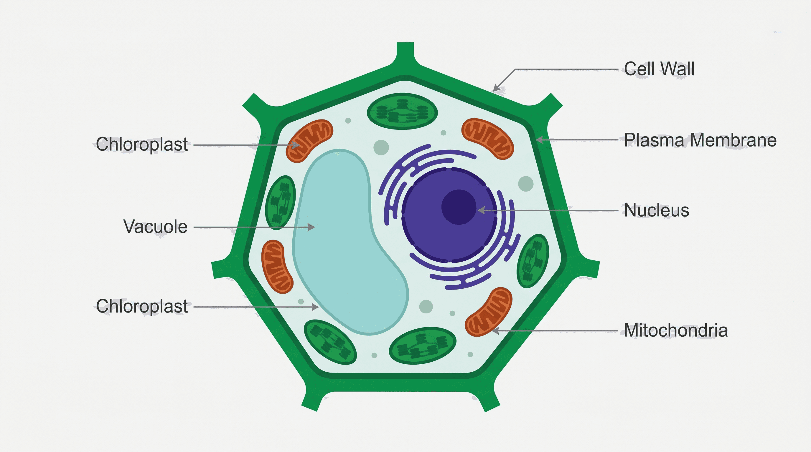
- Drawing one large central vacuole. That is a typical plant-cell feature. Animal cells may contain small vesicles or vacuoles instead.
- Pointing a leader line near an organelle. The end of the line should touch the intended structure, not the surrounding cytoplasm.
- Drawing rough and smooth ER as unrelated objects. They are connected parts of the endomembrane system; rough ER is studded with ribosomes.
- Making every organelle the same size. The nucleus should dominate a typical teaching diagram, while ribosomes are represented as very small dots.
- Using arrows where label lines are intended. Arrows imply movement or a process. Use plain leader lines for names.
- Treating a textbook schematic as a microscope image. A diagram simplifies and separates structures for clarity; it is not a literal scale model.
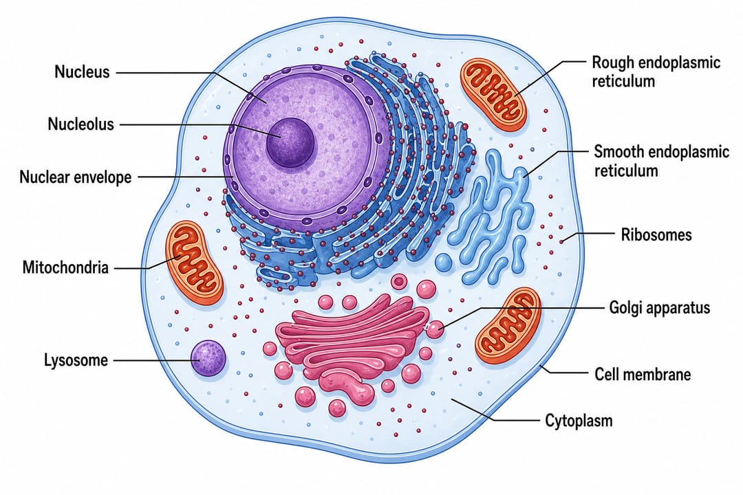
A labeled animal cell diagram should make each organelle easy to identify without crossing leader lines.
Parts of an animal cell and their functions
| Cell part | What it looks like in a diagram | Main function |
|---|---|---|
| Cell membrane | Thin outer boundary | Controls movement into and out of the cell and supports cell signaling |
| Cytoplasm | Open interior surrounding organelles | Contains the cytosol and is the setting for many metabolic reactions |
| Nucleus | Large round or oval compartment | Stores DNA and regulates gene expression |
| Nucleolus | Dense spot inside the nucleus | Produces ribosomal RNA and assembles ribosomal subunits |
| Mitochondrion | Bean-shaped organelle with folded inner membrane | Produces most cellular ATP through aerobic respiration |
| Rough ER | Folded membranes covered with dots | Synthesizes and begins processing secreted and membrane proteins |
| Smooth ER | Tubular membranes without dots | Synthesizes lipids and supports detoxification and calcium storage |
| Golgi apparatus | Stack of curved, flattened sacs | Modifies, sorts, and packages proteins and lipids |
| Ribosomes | Tiny dots, free or attached to rough ER | Translate mRNA into protein |
| Lysosome | Small membrane-bound sphere | Breaks down macromolecules and worn cellular material |
| Peroxisome | Small membrane-bound sphere | Oxidizes fatty acids and detoxifies hydrogen peroxide |
| Centrosome | Pair of centrioles near the nucleus | Organizes microtubules and helps form the mitotic spindle |
| Cytoskeleton | Fine fibers through the cytoplasm | Maintains shape, positions organelles, and supports movement |
| Vesicles | Small membrane bubbles | Transport material between organelles and the cell membrane |
For a middle-school diagram, the first nine entries are usually enough. AP Biology, university, or textbook figures may also label the nuclear envelope, chromatin, peroxisomes, centrosome, and the three main cytoskeletal systems.
How to label an animal cell diagram step by step
1. Find the cell membrane
Start with the thin outside boundary. Label it cell membrane or plasma membrane. Do not add a second thick outer wall.
2. Label the nucleus and nucleolus
The nucleus is normally the largest organelle in a teaching diagram. Point one leader line to the nucleus, another to the nucleolus, and, when required, a third to the nuclear envelope.
3. Separate rough ER from smooth ER
Look for a folded membrane network near the nucleus. The region covered in ribosome dots is rough ER; the region without dots is smooth ER. Give each its own label.
4. Identify the Golgi apparatus
The Golgi is drawn as a curved stack of flattened sacs, often with small transport vesicles nearby. It should not be confused with the ER, which is more continuous and web-like.
5. Add mitochondria
Mitochondria are usually bean-shaped with folds called cristae inside. Label the whole organelle in a basic diagram; label the outer membrane, inner membrane, cristae, and matrix only in a detailed inset.
6. Mark lysosomes, vesicles, and peroxisomes
These can all look like small circles. Use the source diagram or legend rather than guessing. If the illustration does not distinguish them, do not invent an identity.
7. Label ribosomes and cytoplasm last
Point the ribosome label to a visible dot. Point the cytoplasm label into open interior space between organelles. This prevents the two labels from competing for the same area.
8. Check every leader line
Trace each line from its text to its endpoint. Move labels outward, keep text horizontal, and avoid crossings. A clean labeling system is part of the scientific communication, not just decoration.
Labeled vs. unlabeled animal cell diagrams
A labeled animal cell diagram is best for reference, notes, lecture slides, and answer keys. An unlabeled animal cell diagram is best for retrieval practice, quizzes, and worksheets.
To build a matched worksheet set:
- Generate the labeled version first and verify every structure.
- Keep the same cell layout and remove only the text.
- Leave blank callout boxes or numbered leader lines.
- Put the labeled answer key on a separate page.
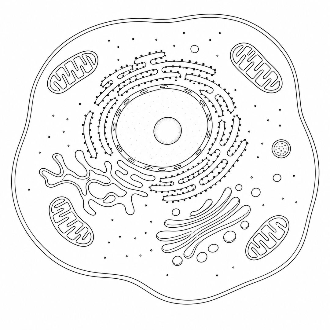
An unlabeled animal cell diagram should preserve the same organelle layout as its answer key.
Animal cell vs. plant cell diagram
| Feature | Animal cell | Plant cell |
|---|---|---|
| Outer boundary | Cell membrane | Cell wall outside a cell membrane |
| Typical shape | Rounded or irregular | More regular and box-like |
| Chloroplasts | Absent | Present in photosynthetic cells |
| Vacuoles | Small vesicles or vacuoles | One large central vacuole is typical |
| Centrosome with centrioles | Common in teaching diagrams | Centrioles absent from most higher plant cells |
| Lysosomal digestion | Lysosomes are prominent | Lytic vacuoles perform many similar roles |
| Shared organelles | Nucleus, mitochondria, ER, Golgi, ribosomes, cytoskeleton | The same shared eukaryotic organelles |
Use the plant vs. animal cell diagram maker for a side-by-side comparison, or read the plant cell labeling guide for plant-specific structures.
A better prompt for an animal cell diagram
A vague prompt leaves too many scientific and layout choices unspecified.
Poor prompt
Draw an animal cell.
Better prompt
Create a clean, labeled animal cell diagram for AP Biology. Show the cell membrane, cytoplasm, nucleus with nucleolus and nuclear envelope, mitochondria with visible cristae, rough and smooth ER, Golgi apparatus, free and bound ribosomes, lysosomes, peroxisomes, centrosome with centrioles, vesicles, and cytoskeleton. Use horizontal English labels outside the cell with thin, non-crossing leader lines. White background, textbook vector style, 16:9.
For a worksheet, add:
Produce a second version with the same layout but no text. Keep blank callout boxes and leader lines so students can label the parts.
Accuracy checklist before you use the diagram
- The cell has a membrane but no cell wall or chloroplasts.
- Every requested organelle appears once or in a biologically plausible number.
- Rough ER carries ribosome dots; smooth ER does not.
- The nucleus contains a nucleolus and is enclosed by a nuclear envelope.
- Mitochondria show inner folds rather than a random spiral.
- Leader lines touch the correct structures and do not cross.
- Labels use consistent singular or plural forms.
- The level of detail matches the class, slide, or publication.
Make an animal cell diagram with SciDraw AI
Open the animal cell diagram generator, choose a labeled, unlabeled, or AP Biology example, and edit the prompt to match your course. For a broader figure that combines cells with pathways or experimental steps, use the cell illustration generator or scientific figure maker.
The generated diagram is a draft, so check every label against your textbook or an authoritative biology source before submitting, teaching, or publishing it.