Gerador Gratuito de Diagrama de Célula Animal
Crie um diagrama de célula animal rotulado a partir de texto simples
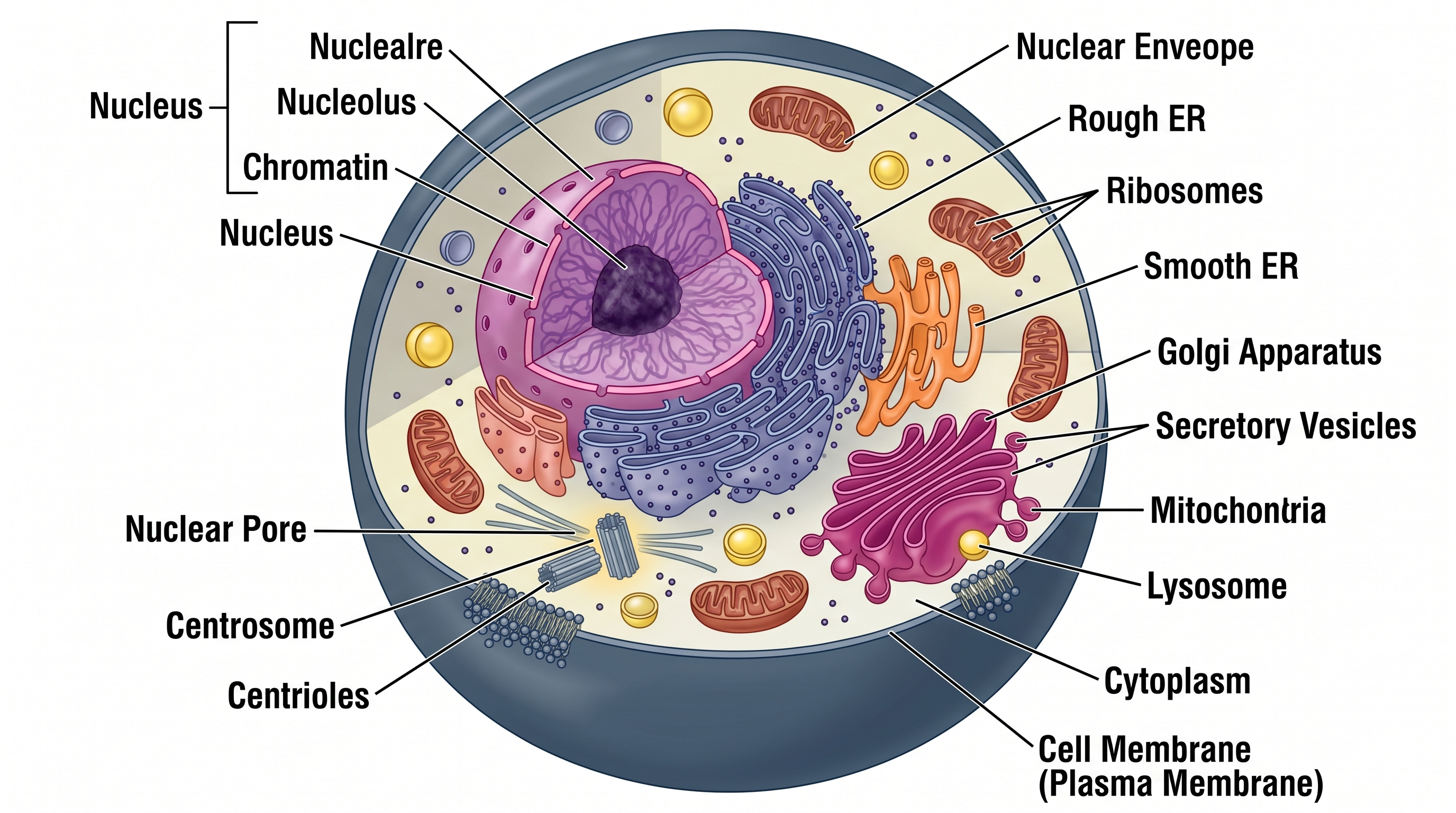
Descreva as organelas e o nível de detalhe necessário, e a IA desenha um diagrama de célula animal limpo e totalmente rotulado — núcleo, mitocôndrias, RE, complexo de Golgi, lisossomo, membrana plasmática e muito mais — pronto para tarefas, slides e publicações.
Exemplos de diagramas de célula animal
Clique em qualquer exemplo para carregar seu prompt ou use-o como ponto de partida para seu próprio diagrama de célula rotulado.
O que faz este gerador de diagrama de célula animal?
Ele converte uma descrição em texto simples de uma célula animal em um diagrama limpo e rotulado mostrando as organelas especificadas. Você descreve quais partes celulares incluir e com que nível de detalhe, e a IA renderiza uma figura cientificamente precisa com rótulos, linhas de chamada e proporções adequadas. Você pode pedir um diagrama de célula animal totalmente rotulado para estudo, ou uma versão em branco, sem legendas, para uma atividade ou quiz em que os alunos preenchem as partes da célula. O resultado é um diagrama organizado e pronto para publicação, sem necessidade de software de ilustração ou desenho manual.
Por que usar um gerador de diagrama de célula animal
- Diagramas de célula animal rotulados são um elemento central dos cursos de biologia, do ensino médio à universidade — os alunos os precisam constantemente para anotações, tarefas e provas.
- Desenhar uma célula detalhada à mão ou em softwares de design genérico é demorado e difícil de revisar quando os requisitos mudam.
- Confira a localização e as proporções das organelas com as referências do curso antes de usar um diagrama para ensinar ou estudar.
- Pesquisadores e educadores precisam de figuras rapidamente personalizáveis para livros didáticos, pôsteres e slides de aula.
- Regenerar a partir de uma descrição revisada é muito mais rápido do que redesenhar ou rerotular uma figura de célula manualmente.
Como criar um diagrama de célula animal
Liste as organelas que deseja incluir e quaisquer detalhes estruturais importantes — por exemplo, «mostre o envelope nuclear com poros, RE rugoso e liso conectados, Golgi com faces cis e trans, e rotule a membrana plasmática como bicamada fosfolipídica». Especifique o propósito do diagrama (tarefa escolar, biologia avançada, livro didático) para que o estilo seja adequado e diga se você quer a figura totalmente rotulada ou deixada sem legendas, como uma atividade em branco para os alunos completarem. Gere o diagrama, verifique se as posições e os rótulos das organelas estão corretos e, em seguida, refine a descrição e regenere até que a figura atenda às suas necessidades.
Partes de uma célula animal
- Membrana plasmática — a bicamada fosfolipídica que envolve a célula e regula o que entra e sai.
- Núcleo — a organela delimitada por membrana que abriga o DNA e dirige as atividades celulares; contém o nucléolo.
- Mitocôndrias — a organela produtora de energia que gera ATP por meio da respiração celular; possui cristas internas.
- Retículo endoplasmático (RE) — o RE rugoso (com ribossomos) sintetiza proteínas; o RE liso sintetiza lipídios e detoxifica.
- Complexo de Golgi — recebe, modifica, empacota e exporta proteínas e lipídios por meio de vesículas.
- Lisossomos — sacos delimitados por membrana contendo enzimas digestivas que degradam resíduos e material estranho.
Perguntas Frequentes sobre o Diagrama de Célula Animal
Quais organelas deve incluir um diagrama de célula animal rotulado?
Um diagrama completo de célula animal tipicamente rotula a membrana plasmática, núcleo (com nucléolo e envelope nuclear), mitocôndrias, retículo endoplasmático rugoso e liso, complexo de Golgi, lisossomos, ribossomos, centrossomo (com centríolos), citoplasma e citoesqueleto. Você pode adicionar vesículas, vacúolos ou peroxissomos dependendo do nível do curso.
Qual é a diferença entre um diagrama de célula animal e de célula vegetal?
As células animais não possuem parede celular, cloroplastos e grande vacúolo central — elementos que aparecem nos diagramas de célula vegetal. As células animais têm centríolos (usados na divisão celular) e lisossomos mais proeminentes do que as células vegetais. Escolher o modelo correto é essencial para a precisão nas tarefas.
Como criar um diagrama de célula animal para biologia avançada?
Descreva todas as organelas abordadas no currículo de biologia avançada — núcleo, mitocôndrias, RE rugoso e liso, complexo de Golgi, ribossomos, lisossomos, centrossomo, membrana plasmática e citoesqueleto. Especifique que os rótulos devem incluir dicas de função, se necessário. Gere, verifique a precisão em relação ao seu livro didático e refine.
Posso rotular as funções das organelas, não apenas os nomes?
Sim — solicite anotações de função no prompt, como «rotule as mitocôndrias como local de produção de ATP» ou «adicione uma nota de que o Golgi modifica e exporta proteínas». A IA incorporará esses elementos nos rótulos ou como legenda.
O diagrama é cientificamente preciso?
O SciDraw AI gera diagramas com base em convenções estabelecidas de ilustração biológica. Sempre compare as posições e proporções das organelas com seu livro didático ou materiais do curso antes de submeter para avaliação.
É gratuito gerar um diagrama de célula animal?
Cada geração usa um pequeno número de créditos. Novas contas recebem créditos gratuitos, então você pode criar um diagrama de célula animal rotulado sem assinatura.
Qual é a diferença entre um diagrama de célula animal rotulado e um sem legendas?
Um diagrama de célula animal rotulado nomeia cada organela com linhas de chamada, sendo ideal para estudo e consulta. Um diagrama de célula animal sem legendas (em branco) mostra os mesmos contornos de organelas sem texto, deixando espaço para preencher os nomes — perfeito para revisão, autoavaliação ou impressão como atividade. Basta dizer qual versão você quer ao gerar, e você pode alternar entre elas a qualquer momento.
Posso criar uma atividade de diagrama de célula animal ou uma tarefa de 'rotular as partes da célula'?
Sim. Peça um diagrama de célula animal sem legendas ou em branco e o gerador desenha contornos claros de organelas sem texto, pronto para imprimir como uma atividade ou quiz sobre as partes da célula. Você também pode gerar um gabarito rotulado correspondente mostrando cada parte da célula animal e seu nome, para que os alunos confiram após rotular.
Escrevendo um artigo, TCC, tese ou projeto de pesquisa?
O SciDraw AI também cria figuras de pesquisa prontas para publicar: resumos gráficos, figuras de mecanismo, fluxogramas e modelos conceituais.
Explore mais ferramentas
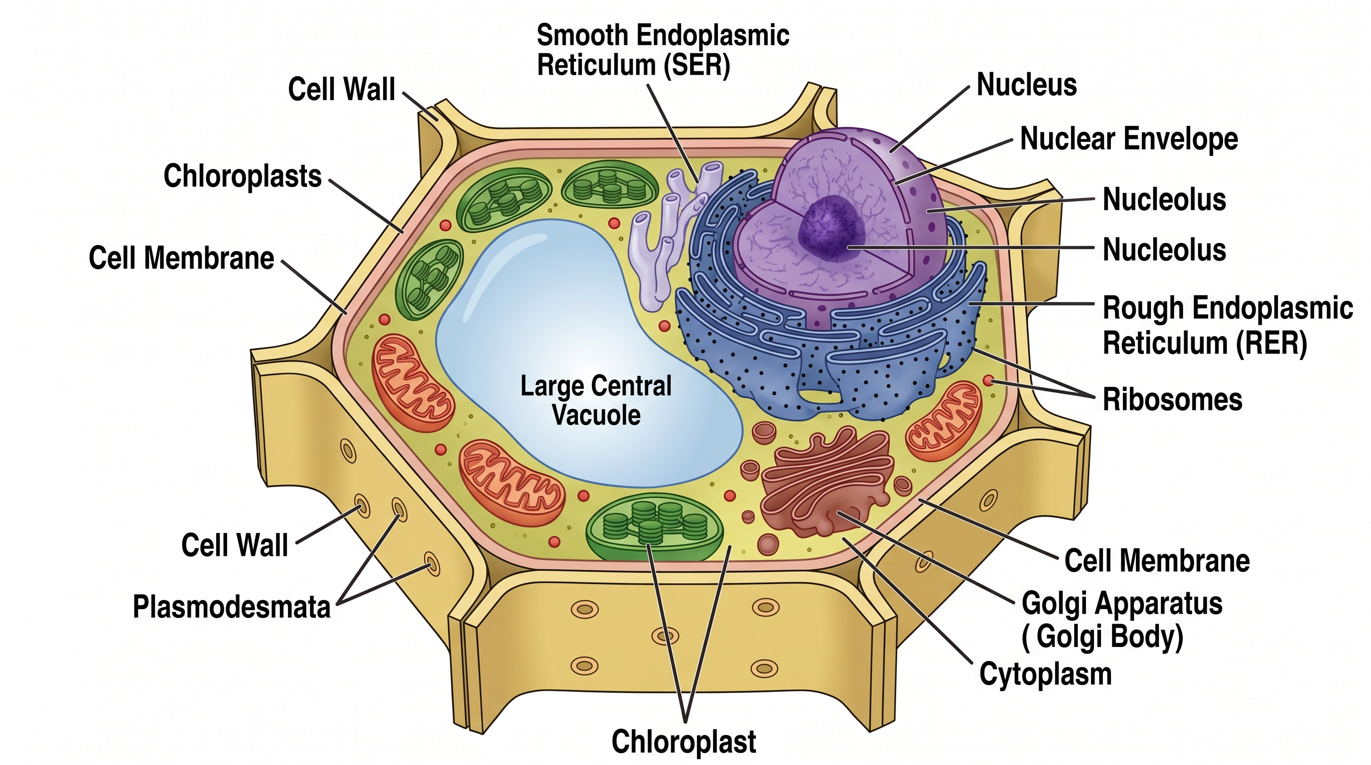
Diagrama de Célula Vegetal
Gere um diagrama de célula vegetal rotulado com todas as organelas.
Experimente grátis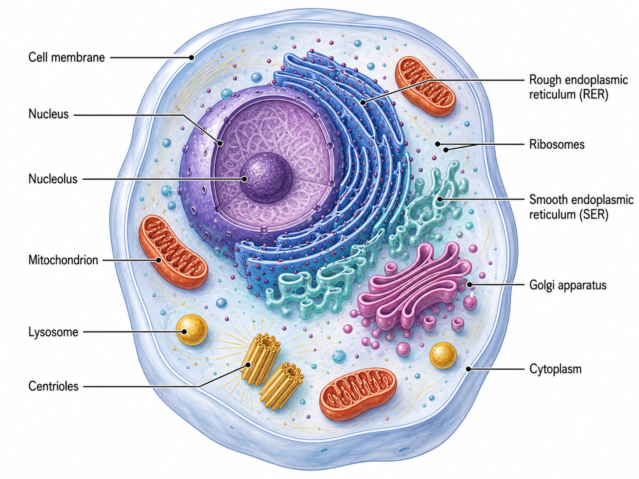
Gerador de Ilustrações Celulares
Crie diagramas de biologia celular, organelas e processos celulares
Experimente grátis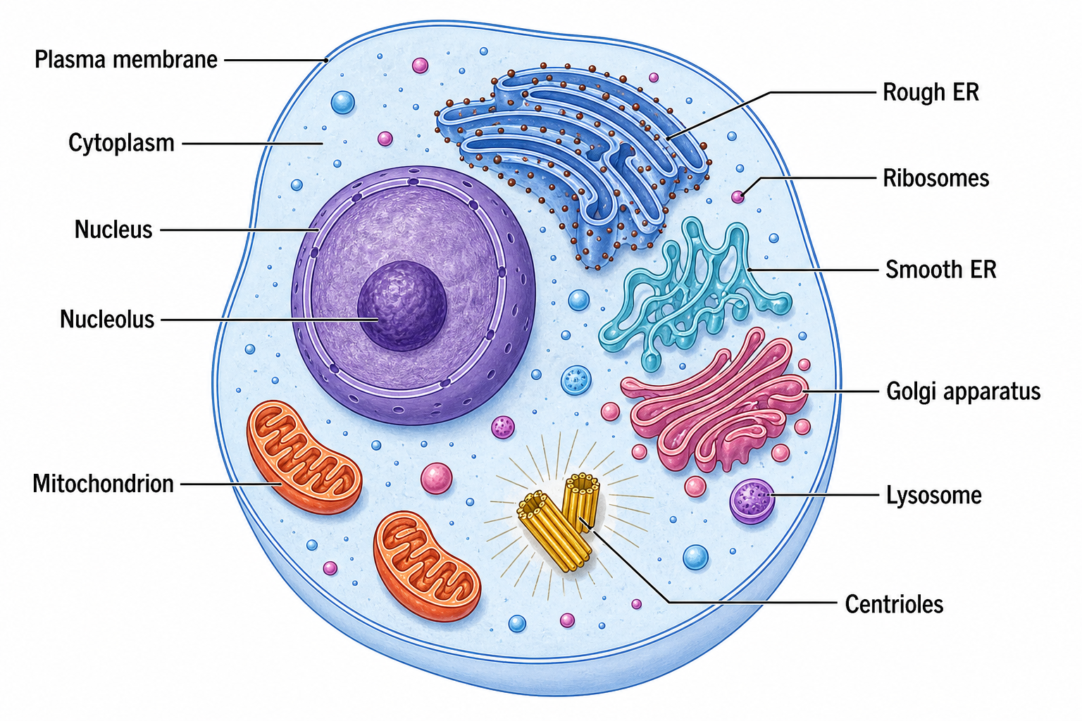
Gerador de Diagrama de Célula
Desenhe células animais, vegetais, eucariontes e procariontes legendadas, com as organelas corretas.
Experimente grátisPrecisa de outros diagramas de célula ou biologia?
Gere diagramas de célula vegetal, fases da mitose, cladogramas, teias alimentares e muito mais com o SciDraw AI.