Plant cell diagrams show up in almost every biology textbook, exam, and lab report — yet labeling one correctly trips up students at every level. Get a single organelle wrong and the whole diagram loses marks. This guide walks you through every part of a plant cell, explains what each organelle does, and shows you practical tips for producing accurate, professional-looking labeled diagrams.
What you'll learn in this guide:
- The name and function of every major plant cell organelle
- A quick-reference table of organelles and their roles
- Key differences between plant and animal cells
- Step-by-step labeling strategies for exams and reports
- How to create and label a plant cell diagram with SciDraw AI
Need a labeled diagram right now? The SciDraw AI plant cell diagram generator draws a fully labeled plant cell — cell wall, chloroplasts, vacuole, and every organelle — from a single sentence. Use this guide to learn the parts, then generate a clean, exam-ready version in seconds.
Why Labeling Matters
A diagram without labels is just a picture. In science, labels communicate precise meaning — they tell the reader which structure performs which function, and they form the foundation of every explanation you write. Whether you are preparing for an AP Biology test, writing a university lab report, or teaching a class, a well-labeled plant cell diagram shows that you understand how the cell works, not just what it looks like.
The Full Plant Cell: An Overview
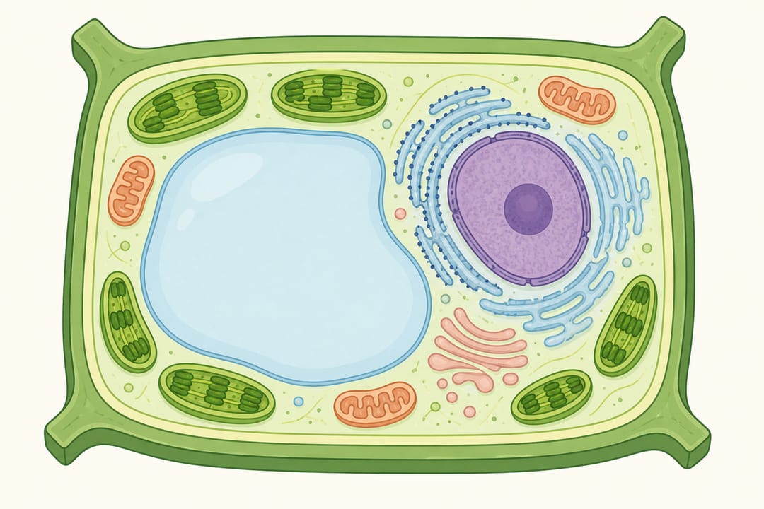
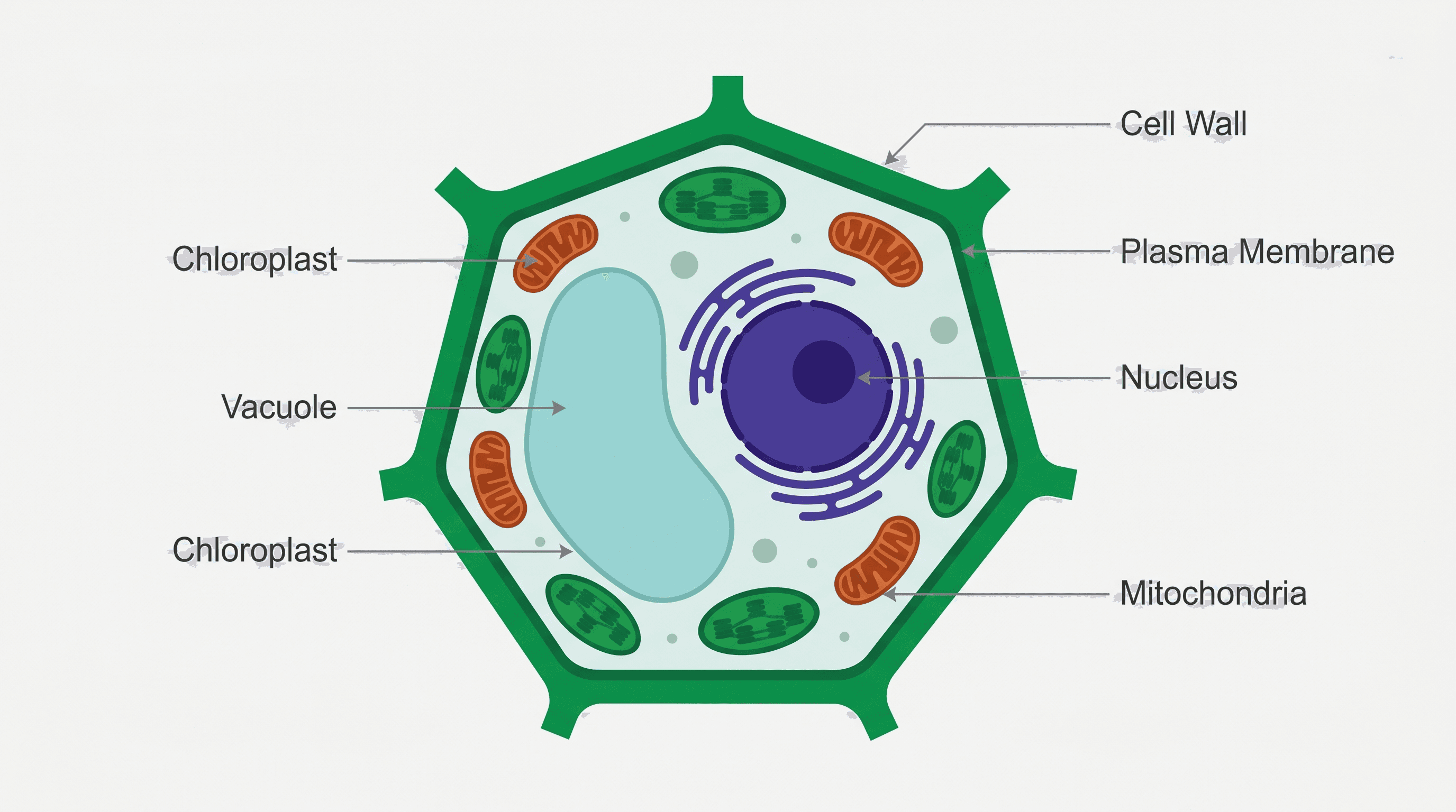
Plant cells are eukaryotic cells — they have a membrane-bound nucleus and a range of specialized organelles. Compared with animal cells, they have three unique structures: the cell wall, the chloroplast, and the central vacuole. These additions allow plants to photosynthesize, maintain rigidity without a skeleton, and store large quantities of water and nutrients.
A typical plant cell diagram includes around 10–14 labeled components, depending on the level of detail required.
A typical plant cell in cross-section, ready to be labeled with SciDraw AI.
Labeled vs. Blank Plant Cell Diagrams
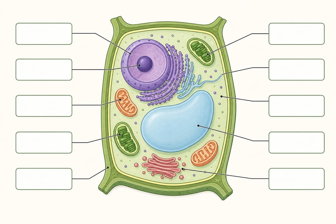
Most study workflows need both versions of the same figure. A labeled plant cell diagram names every organelle for reference and revision, while a blank (unlabeled) version turns the same picture into a worksheet you can fill in from memory.
- Labeled plant cell diagram — every structure is named with a clean leader line. Use it as an answer key, a revision poster, or a figure in a lab report.
- Blank plant cell diagram — the same layout with the labels removed and the leader lines left in place. Use it to test recall before an exam.
- Partially labeled — a few structures named as anchors (nucleus, cell wall, chloroplast) and the rest left for the student. Ideal for guided practice.
The fastest way to get a plant cell labeled correctly is to generate the labeled version first and verify it against the organelle list below, then generate a blank plant cell worksheet as a second figure. Ask the plant cell diagram generator for the same view and the same organelles in both prompts so the worksheet and the answer key line up for your students.
Plant Cell Organelles and Their Functions
1. Cell Wall
The cell wall is the outermost rigid layer, composed primarily of cellulose fibers. It gives the cell a fixed shape, prevents over-expansion when the cell absorbs water, and provides structural support to the entire plant. Unlike the cell membrane, the cell wall is fully permeable — it does not control what enters or exits.
Labeling tip: Draw the cell wall as a thick, uniform rectangle around the outside of the cell. It is distinct from the much thinner cell membrane just inside it.
2. Cell Membrane (Plasma Membrane)
Just inside the cell wall sits the cell membrane, a thin, flexible phospholipid bilayer that controls the movement of substances into and out of the cell. It is selectively permeable, allowing small molecules like water and oxygen through while blocking larger or charged particles.
Labeling tip: Use a fine line immediately inside the cell wall. Label both structures separately — examiners often deduct marks when students conflate them.
3. Cytoplasm
The cytoplasm is the gel-like fluid (cytosol) that fills the cell interior, surrounding all organelles. It is the medium in which metabolic reactions take place and through which molecules diffuse from one organelle to another.
Labeling tip: You cannot point to cytoplasm as a discrete object. Use a leader line into the open space of the cell interior.
4. Nucleus
The nucleus is the control center of the cell. It contains the cell's genetic material (DNA) in the form of chromosomes, and it coordinates cell activities including growth, metabolism, protein synthesis, and reproduction.
Inside the nucleus, label:
- Nuclear envelope — double membrane surrounding the nucleus
- Nucleolus — dense region where ribosomal RNA is synthesized
- Chromatin/chromosomes — DNA and associated proteins
Labeling tip: Draw the nucleus as a large oval, not a perfect circle, and show the double membrane clearly.
5. Chloroplast
Chloroplasts are the defining organelles of plant cells — they are where photosynthesis occurs. Chloroplasts contain a green pigment called chlorophyll, which absorbs light energy to convert carbon dioxide and water into glucose and oxygen.
Internally, chloroplasts have:
- Thylakoids — flattened membrane sacs where the light-dependent reactions occur
- Grana — stacks of thylakoids
- Stroma — fluid surrounding the grana where the Calvin cycle (light-independent reactions) takes place
Labeling tip: Draw chloroplasts as green, oval, double-membraned structures, typically near the outer edges of the cell where light penetrates.
6. Central Vacuole
Plant cells have a single, large central vacuole that can occupy up to 90% of the cell's volume in mature cells. It stores water, ions, sugars, pigments, and waste products. When the vacuole is full, it presses against the cell wall, creating turgor pressure — the force that keeps non-woody plant tissue firm.
Labeling tip: Draw the central vacuole as a large, central space bounded by the tonoplast (vacuolar membrane). In mature cells it dominates the diagram.
7. Mitochondria
Mitochondria (singular: mitochondrion) are the sites of aerobic respiration — the process that converts glucose and oxygen into ATP (adenosine triphosphate), the cell's primary energy currency. Although plants can make glucose through photosynthesis, they still need mitochondria to release the chemical energy stored in that glucose.
Key internal features:
- Outer membrane — smooth boundary
- Inner membrane — folded into cristae to maximize surface area
- Matrix — fluid interior where the Krebs cycle occurs
Labeling tip: Draw mitochondria as small, bean-shaped structures with internal folded membranes (cristae). They are smaller than chloroplasts.
8. Endoplasmic Reticulum (ER)
The endoplasmic reticulum is a network of interconnected membrane-bound tubules and sacs. There are two types:
- Rough ER (RER) — studded with ribosomes; involved in synthesizing and folding proteins that will be secreted or sent to other organelles
- Smooth ER (SER) — lacks ribosomes; involved in lipid synthesis, carbohydrate metabolism, and detoxification
Labeling tip: Show the ER as a series of wavy, parallel membranes connected to the nuclear envelope.
9. Golgi Apparatus (Golgi Body)
The Golgi apparatus is the cell's "post office." It receives proteins from the rough ER, modifies and packages them, and ships them to their final destinations — either secreted from the cell or sent to organelles like the vacuole or cell membrane.
Labeling tip: Draw it as a stack of slightly curved, flattened sacs (cisternae), distinct from the ER. Label the cis face (receiving) and trans face (shipping) if detail is required.
10. Ribosomes
Ribosomes are the molecular machines that synthesize proteins by translating messenger RNA (mRNA). They are found:
- On the surface of rough ER
- Free in the cytoplasm
- Inside chloroplasts and mitochondria (prokaryote-like ribosomes, supporting the endosymbiotic theory)
Labeling tip: Ribosomes are very small — represent them as tiny dots on the rough ER or scattered in the cytoplasm.
11. Plasmodesmata
Plasmodesmata are microscopic channels that pass through the cell walls of adjacent plant cells, allowing direct communication and transport between cells. They are unique to plant cells and form a continuous cytoplasmic network called the symplast.
Labeling tip: Show plasmodesmata as thin lines piercing the cell wall at intervals.
12. Amyloplasts (Optional Detail)
Amyloplasts are a type of plastid that stores starch granules. They are especially prominent in root and storage cells. They are not always required in standard diagrams, but including them in a root cell diagram is accurate.
Quick-Reference Table: Plant Cell Organelles and Functions
| Organelle | Membrane | Key Function | Unique to Plants? |
|---|---|---|---|
| Cell wall | None (non-living) | Structural support, shape | Yes |
| Cell membrane | Single | Selectively permeable barrier | No |
| Nucleus | Double (envelope) | Gene storage, cell control | No |
| Chloroplast | Double | Photosynthesis | Yes |
| Central vacuole | Single (tonoplast) | Water/nutrient storage, turgor pressure | Yes (large) |
| Mitochondrion | Double | Aerobic respiration, ATP production | No |
| Rough ER | Single | Protein synthesis and transport | No |
| Smooth ER | Single | Lipid synthesis, detoxification | No |
| Golgi apparatus | Single | Protein modification and packaging | No |
| Ribosomes | None | Protein synthesis | No |
| Plasmodesmata | Continuous with membrane | Cell-to-cell communication | Yes |
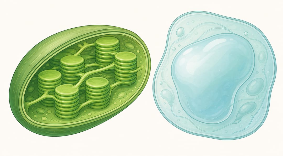
Inside the chloroplast and central vacuole — organelles unique to plant cells.
Plant Cell vs. Animal Cell: Key Differences
Understanding what makes plant cells unique helps you label them more accurately — and avoid adding structures that do not belong.
| Feature | Plant Cell | Animal Cell |
|---|---|---|
| Cell wall | Present (cellulose) | Absent |
| Chloroplasts | Present | Absent |
| Central vacuole | Large, single | Small or absent |
| Centrioles | Absent (in most) | Present |
| Shape | Regular, rectangular | Irregular, rounded |
| Plasmodesmata | Present | Absent |
| Lysosomes | Rare | Common |
For the animal-cell counterpart, read our animal cell labeling guide, then use the animal cell diagram generator to make a labeled, unlabeled, or AP Biology version.
Step-by-Step: How to Label a Plant Cell Diagram
Step 1 — Start with the outer boundary
Label the cell wall first (outermost), then the cell membrane just inside it. These two are frequently confused and examiners look carefully at which is which.
Step 2 — Identify the nucleus
The nucleus is typically the largest, most prominent organelle. Label the nuclear envelope, nucleolus, and chromatin. Draw leader lines clearly so they do not cross.
Step 3 — Add the chloroplasts
Place 4–6 chloroplasts near the outer edges. Label the outer/inner membrane, thylakoids, and stroma if required.
Step 4 — Mark the central vacuole
In a mature cell, the central vacuole takes up most of the interior. Label the vacuole and the tonoplast (surrounding membrane).
Step 5 — Add mitochondria and ER
Scatter 2–3 mitochondria in the remaining cytoplasm. Add the rough and smooth ER connecting to the nuclear envelope.
Step 6 — Include the Golgi apparatus
Draw the Golgi near the nucleus or ER. Connect vesicles budding from the trans face.
Step 7 — Dot in ribosomes
Add small dots on the rough ER surface and free in the cytoplasm.
Step 8 — Review and cross-check
Check every label with the table above. Make sure leader lines touch the correct structure, labels are horizontal (not diagonal), and every label is spelled correctly.
Position clear leader lines first, then add each label as you cross-check the diagram.
Common Labeling Mistakes to Avoid
- Confusing the cell wall and cell membrane — the wall is outermost and rigid; the membrane is inner and flexible.
- Omitting the tonoplast — the vacuole membrane is a separate structure from the vacuole itself.
- Drawing centrioles in a plant cell — most plant cells do not have centrioles; this is an animal cell feature.
- Merging the ER with the nuclear envelope — they are continuous, but label them separately.
- Using the wrong scale — the central vacuole should be much larger than individual organelles like mitochondria.
Creating a Labeled Plant Cell Diagram with SciDraw AI
Drawing a plant cell by hand takes time and artistic skill — and hand-drawn diagrams can be hard to edit or reuse. SciDraw AI's cell illustration generator lets you describe the cell type and level of detail you need, and the tool generates a clean, scientifically accurate diagram you can annotate, export, and drop into your lab report or slide deck in minutes.
You can also start from an existing plant cell diagram template and add or remove labels to match your curriculum requirements.
FAQ
Q: How many organelles should I label in a plant cell diagram? A: For GCSE or high-school level, label at least 8–10: cell wall, cell membrane, cytoplasm, nucleus, chloroplast, vacuole, mitochondria, ribosomes, ER, and Golgi. University diagrams often require additional detail such as the tonoplast, plasmodesmata, and internal chloroplast structures.
Q: What is the difference between cell wall and cell membrane? A: The cell wall is the rigid outer layer made of cellulose that provides structural support. The cell membrane (plasma membrane) is the thin, flexible, selectively permeable layer just inside the cell wall that regulates what enters and leaves the cell.
Q: Do plant cells have mitochondria if they already have chloroplasts? A: Yes. Chloroplasts produce glucose through photosynthesis, but mitochondria are still needed to convert that glucose into ATP (usable energy) through cellular respiration. Plant cells require energy at night when photosynthesis is not occurring.
Q: Why is the central vacuole so large in plant cells? A: The central vacuole stores water, maintaining turgor pressure that keeps plant cells firm. It also stores nutrients, waste products, and pigments. As the plant matures, the vacuole expands significantly, often pushing other organelles to the periphery of the cell.
Q: Can I use SciDraw AI to create diagrams for textbooks or publications? A: Yes. SciDraw AI generates publication-quality scientific figures. The cell illustration generator produces vector-quality outputs suitable for research papers, educational materials, and presentations.
Q: What is the function of plasmodesmata? A: Plasmodesmata are narrow channels through plant cell walls that connect adjacent cells. They allow direct transport of water, nutrients, signaling molecules, and even viruses between cells, forming a continuous network called the symplast.