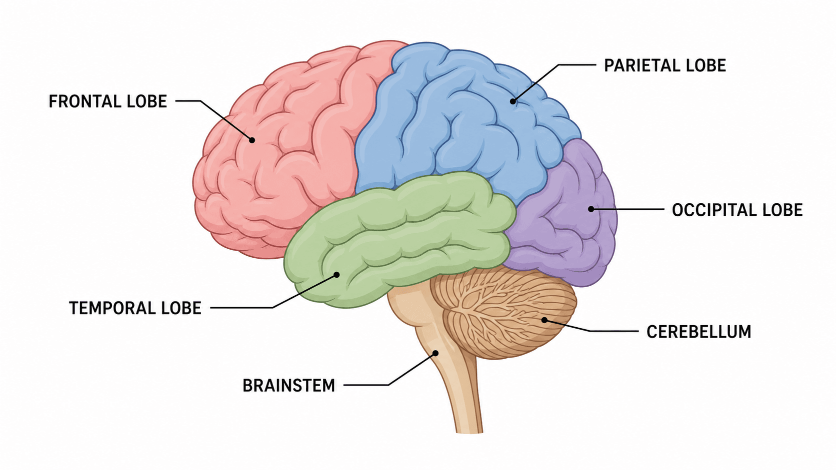
Um diagrama do cérebro pode parecer convincente e mesmo assim perder pontos se os lobos estiverem na posição errada ou se o cerebelo e o tronco encefálico forem trocados. O caminho mais seguro é orientar o desenho primeiro — a frente do cérebro à esquerda —, depois rotular os quatro lobos e só então acrescentar o cerebelo, o tronco encefálico e as estruturas internas.
Este guia explica as partes do cérebro, o que cada região faz e como produzir um diagrama rotulado com precisão. Se você precisa de uma figura pronta agora, o gerador de diagrama do cérebro desenha uma vista lateral rotulada, os quatro lobos ou um corte transversal a partir de uma descrição em linguagem simples.
Erros comuns no diagrama do cérebro
Corrija-os antes de adicionar cor ou sombreamento:
- Colocar o lobo frontal atrás. Em uma vista lateral (de perfil) padrão, a frente do cérebro fica voltada para a esquerda, então o lobo frontal fica à frente, à esquerda.
- Confundir o cerebelo com o tronco encefálico. O cerebelo é a estrutura enrugada na parte inferior de trás; o tronco encefálico fica abaixo e à frente dele, conectando-se à medula espinhal.
- Juntar os lobos occipital e parietal. São regiões distintas: o occipital bem atrás e o parietal acima e à frente dele.
- Rotular o cérebro como um único lobo. O cérebro (telencéfalo) é toda a parte superior, dividida em quatro lobos por hemisfério.
- Traçar linhas de chamada que se cruzam. Mantenha-as curtas e paralelas sempre que possível, para que cada uma aponte claramente para uma região.
- Esquecer o corpo caloso em um corte. Em uma vista sagital mediana, é a faixa que conecta os dois hemisférios, e costuma ser omitida.
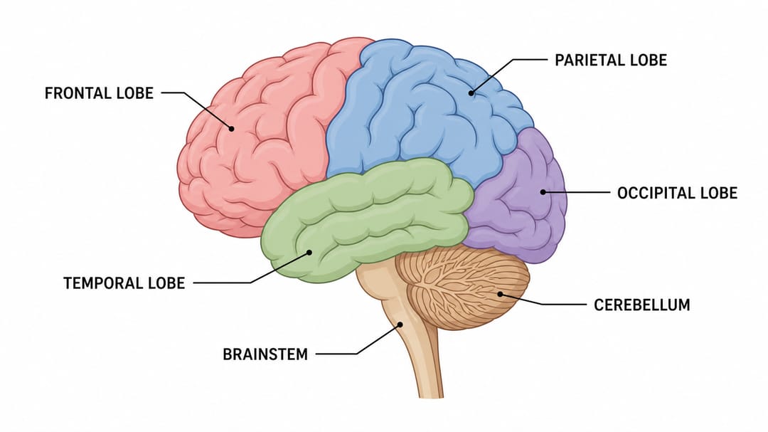
Um diagrama claro do cérebro rotula os quatro lobos mais o cerebelo e o tronco encefálico sem cruzar as linhas de chamada.
Os quatro lobos e suas funções
A maioria dos diagramas do cérebro começa pelos quatro lobos do cérebro.
| Lobo | Localização | Principais funções |
|---|---|---|
| Frontal | Na frente | Tomada de decisões, movimento, fala (área de Broca), personalidade |
| Parietal | No alto, ao centro | Tato, temperatura, noção espacial |
| Temporal | Nas laterais, perto das têmporas | Audição, memória, compreensão da linguagem (área de Wernicke) |
| Occipital | Atrás | Processamento da visão |
Outras estruturas importantes
Além dos lobos, um diagrama completo do cérebro costuma rotular:
- Cerebelo — coordena o movimento e o equilíbrio; é a estrutura enrugada na parte inferior de trás.
- Tronco encefálico — controla funções automáticas vitais, como respiração e frequência cardíaca; conecta o cérebro à medula espinhal. Inclui o mesencéfalo, a ponte e o bulbo.
- Corpo caloso — a faixa de fibras que liga os hemisférios esquerdo e direito (visível em corte).
- Tálamo e hipotálamo — centros de retransmissão e regulação, mostrados em uma vista sagital mediana.
Passo a passo: rotular o diagrama
- Oriente o cérebro. Confirme que a frente está voltada para a esquerda em uma vista lateral.
- Delimite os quatro lobos. Rotule o frontal, o parietal, o temporal e o occipital em suas posições.
- Adicione o cerebelo na parte inferior de trás.
- Adicione o tronco encefálico abaixo do cerebelo, seguindo em direção à medula espinhal.
- Inclua estruturas internas (corpo caloso, tálamo) apenas se estiver desenhando um corte transversal.
- Confira as linhas de chamada para que nenhuma se cruze e cada uma toque uma região.
Melhores prompts para um diagrama do cérebro com IA
Descreva a vista e os rótulos de que você precisa:
- "Vista lateral com o lobo frontal à frente, à esquerda; rotule os quatro lobos."
- "Inclua o cerebelo e o tronco encefálico como estruturas rotuladas separadamente."
- "Use um corte sagital mediano e adicione o corpo caloso e o tálamo."
- "Mantenha um estilo limpo, de livro didático, com linhas de chamada que não se cruzam."
O gerador de diagrama do cérebro lê essas instruções e produz uma figura editável, para você ajustar o limite de um lobo ou um rótulo com palavras, em vez de redesenhar.
Checklist de precisão
Antes de entregar um diagrama do cérebro, confirme:
- Os quatro lobos estão nas posições corretas.
- O lobo frontal está à frente (à esquerda em uma vista lateral padrão).
- O cerebelo e o tronco encefálico estão rotulados separadamente.
- Todo corte transversal inclui o corpo caloso.
- Cada linha de chamada toca uma região e nenhuma se cruza.
- A vista (lateral ou sagital mediana) corresponde às estruturas mostradas.
Guias de diagramas relacionados
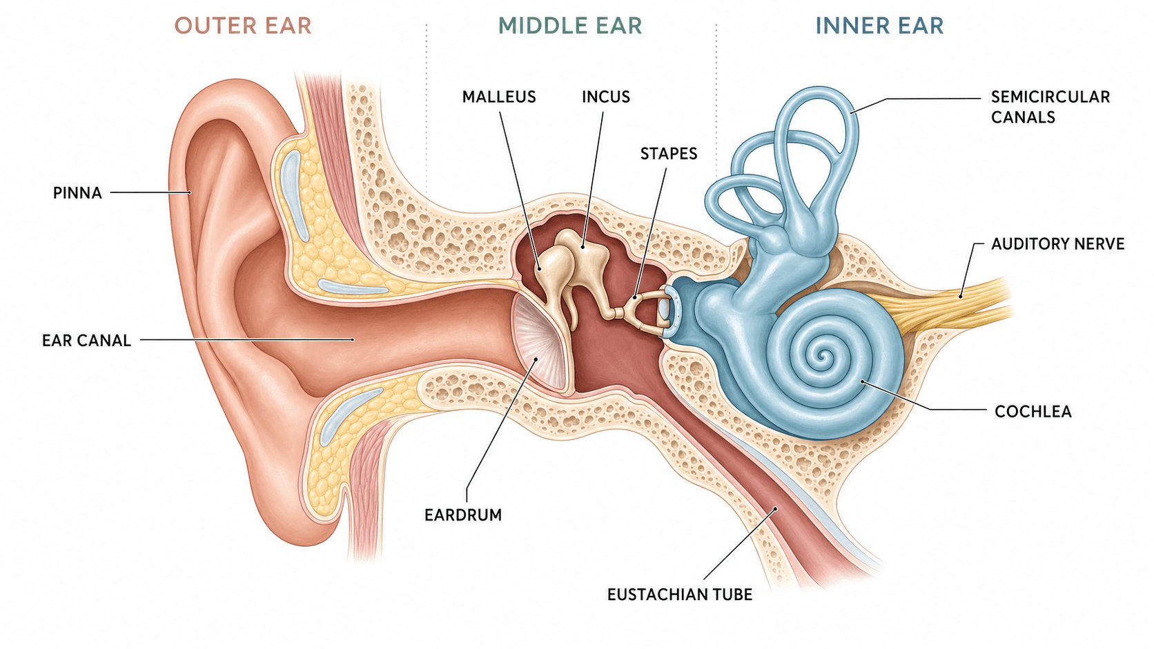
- Como rotular um diagrama do ouvido para outro passo a passo de anatomia.
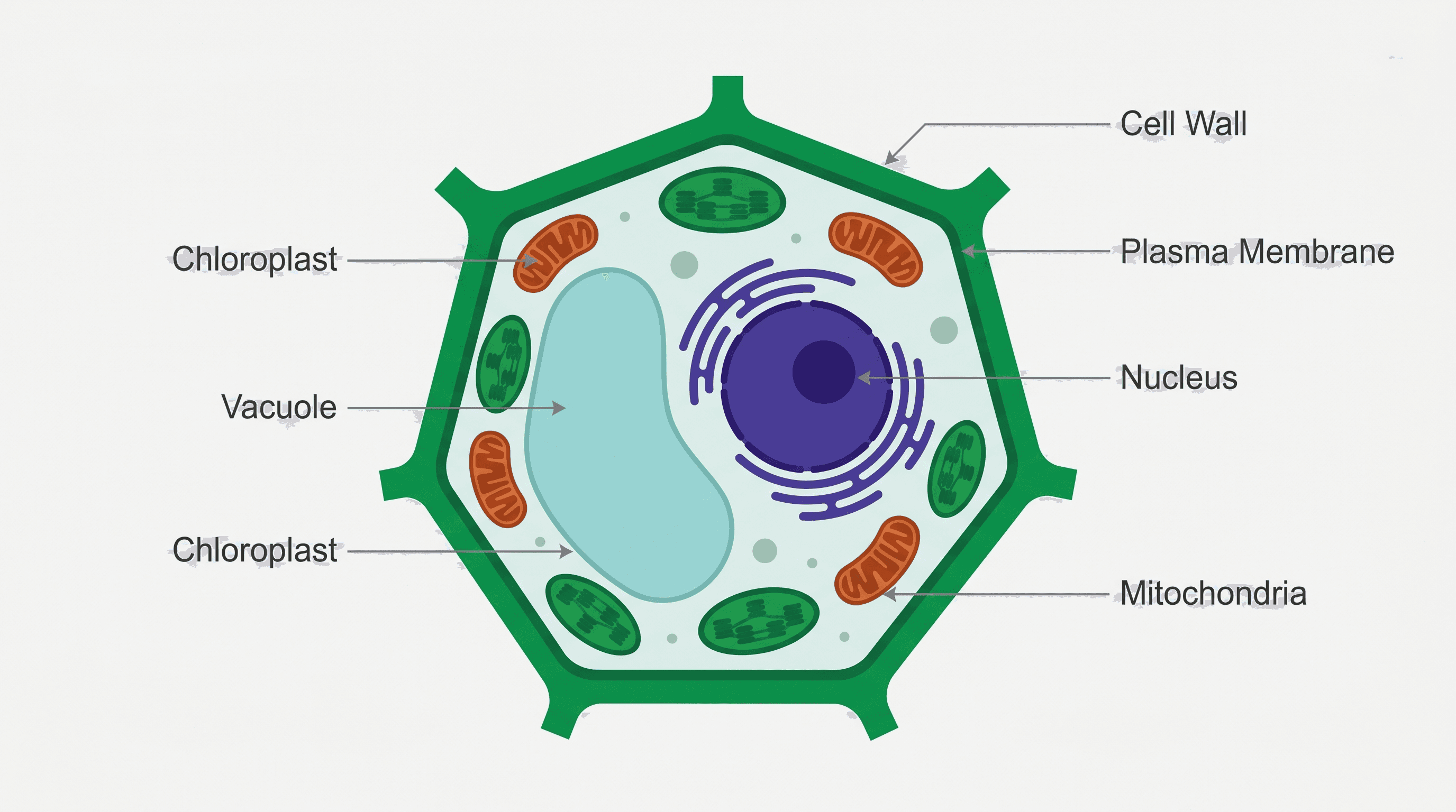
- Como rotular um diagrama de célula animal para figuras de biologia celular.
- Explore mais ferramentas de anatomia, como os geradores de diagrama do sistema nervoso e diagrama do neurônio.
Perguntas frequentes
Quais são os quatro lobos do cérebro? O frontal, o parietal, o temporal e o occipital. Cada hemisfério do cérebro contém os quatro.
Qual é a diferença entre o cerebelo e o tronco encefálico? O cerebelo coordena o movimento e o equilíbrio e fica na parte inferior de trás do cérebro. O tronco encefálico controla funções automáticas, como a respiração, e conecta o cérebro à medula espinhal.
Qual lobo controla a visão? O lobo occipital, na parte de trás do cérebro, processa as informações visuais.
Como faço um diagrama do cérebro rotulado rapidamente? Descreva a vista e as estruturas ao gerador de diagrama do cérebro e exporte uma figura rotulada; depois remova os rótulos para ter uma versão de estudo em branco.