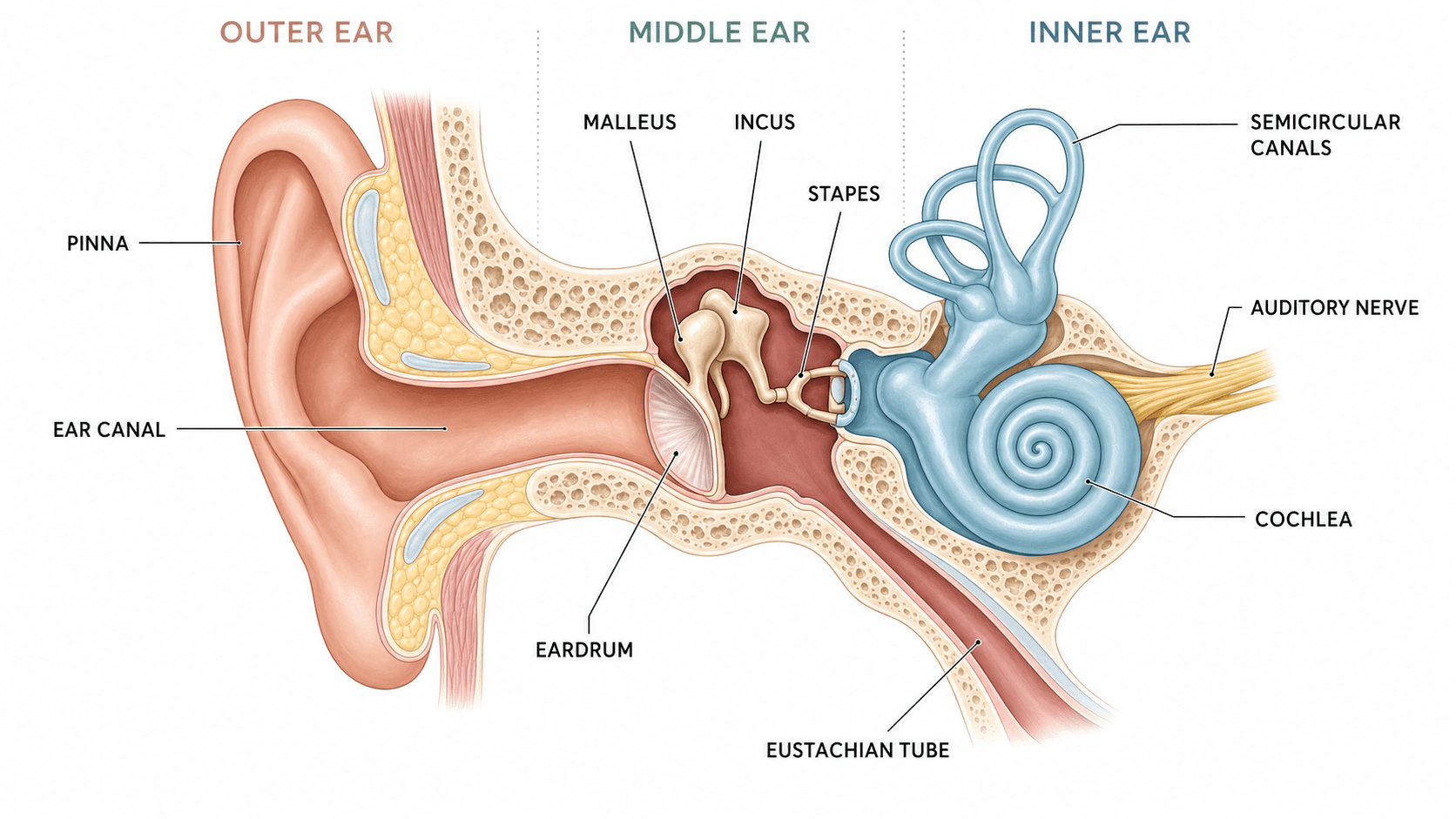
An ear diagram packs a lot of small structures into three regions, and it is easy to place a label in the wrong section or point a leader line at the space between two parts. The dependable method is to divide the ear into its three regions first — outer, middle, and inner — then label the structures within each region in the order sound travels through them.
This guide explains the parts of the ear, what each one does, and how to produce an accurate labeled diagram. If you need a finished figure immediately, the ear anatomy diagram generator can draw a labeled or blank version from a plain-language description.
Common ear diagram mistakes
Fix these before adding color or decoration:
- Confusing the pinna with the whole outer ear. The pinna (auricle) is the visible flap; the outer ear also includes the ear canal.
- Mislabeling the eardrum. The tympanic membrane (eardrum) sits at the boundary between the outer and middle ear, not inside the inner ear.
- Swapping the three ossicles. The order from eardrum inward is malleus (hammer), incus (anvil), stapes (stirrup). The stapes connects to the oval window.
- Placing the cochlea in the middle ear. The cochlea is an inner-ear structure, along with the semicircular canals and the auditory nerve.
- Forgetting the Eustachian tube. It links the middle ear to the throat and equalizes pressure — a commonly omitted label.
- Pointing a leader line at empty space. The end of each line should touch the structure it names, not the gap beside it.
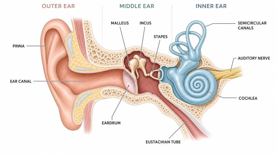
A clear ear diagram separates the outer, middle, and inner ear so each structure is easy to identify.
Parts of the ear by region
Group the labels by region and the diagram becomes far easier to read.
| Region | Structures | Main function |
|---|---|---|
| Outer ear | Pinna (auricle), ear canal | Collects and channels sound waves |
| Middle ear | Eardrum (tympanic membrane), malleus, incus, stapes, Eustachian tube | Amplifies vibrations and equalizes pressure |
| Inner ear | Cochlea, semicircular canals, oval window, auditory (vestibulocochlear) nerve | Converts vibration to nerve signals; balance |
How hearing works (label in this order)
Following the path of sound gives you the correct labeling sequence:
- Pinna collects sound waves and funnels them into the ear canal.
- Sound vibrates the eardrum.
- The three ossicles — malleus, incus, stapes — amplify and transmit the vibration.
- The stapes pushes on the oval window, sending waves into the fluid-filled cochlea.
- Hair cells in the cochlea convert the waves to electrical signals.
- The auditory nerve carries the signal to the brain.
- The semicircular canals handle balance, separate from hearing.
Better prompts for an AI ear diagram
Specific instructions produce accurate figures:
- "Divide the diagram into outer, middle, and inner ear with a subtle region label for each."
- "Label the three ossicles in order: malleus, incus, stapes."
- "Include the Eustachian tube and the auditory nerve."
- "Keep leader lines from crossing and use a clean, textbook style."
The ear anatomy diagram generator turns these into an editable figure, so you can rename a structure or add a label with words instead of redrawing.
Accuracy checklist
Before you submit an ear diagram, confirm:
- Outer, middle, and inner ear are clearly separated.
- The eardrum sits between the outer and middle ear.
- The ossicles are labeled in order: malleus, incus, stapes.
- The cochlea and semicircular canals are in the inner ear.
- The Eustachian tube and auditory nerve are labeled.
- Every leader line touches the structure it names.
Related diagram guides
- How to label an animal cell diagram for cell biology figures.
- How to label a brain diagram for another anatomy walkthrough.
- Explore more anatomy tools like the eye anatomy diagram and heart diagram makers.
FAQ
What are the three main parts of the ear? The outer ear (pinna and ear canal), the middle ear (eardrum and three ossicles), and the inner ear (cochlea, semicircular canals, and auditory nerve).
What are the three bones in the ear called? The malleus (hammer), incus (anvil), and stapes (stirrup). They are the smallest bones in the human body.
Where is the cochlea located? In the inner ear. It converts sound vibrations into electrical signals that the auditory nerve carries to the brain.
How do I make a labeled ear diagram quickly? Describe the regions and structures to the ear anatomy diagram generator and export a labeled figure, then run a second prompt asking for the same view with no labels to get an ear diagram to label.