Eye Anatomy Diagram Generator
Clearly labeled human eye diagrams in seconds
Type what you need and SciDraw AI draws a clean, anatomically reasonable eye diagram with tidy callout labels. Perfect for cross sections, the retina, eye muscles, and the full eyeball structure for biology, optometry, and nursing study. Always verify details against an authoritative source.
Eye diagram examples
Start from a ready-made prompt and adapt it to your lesson, lab, or study notes.
What is an eye anatomy diagram?
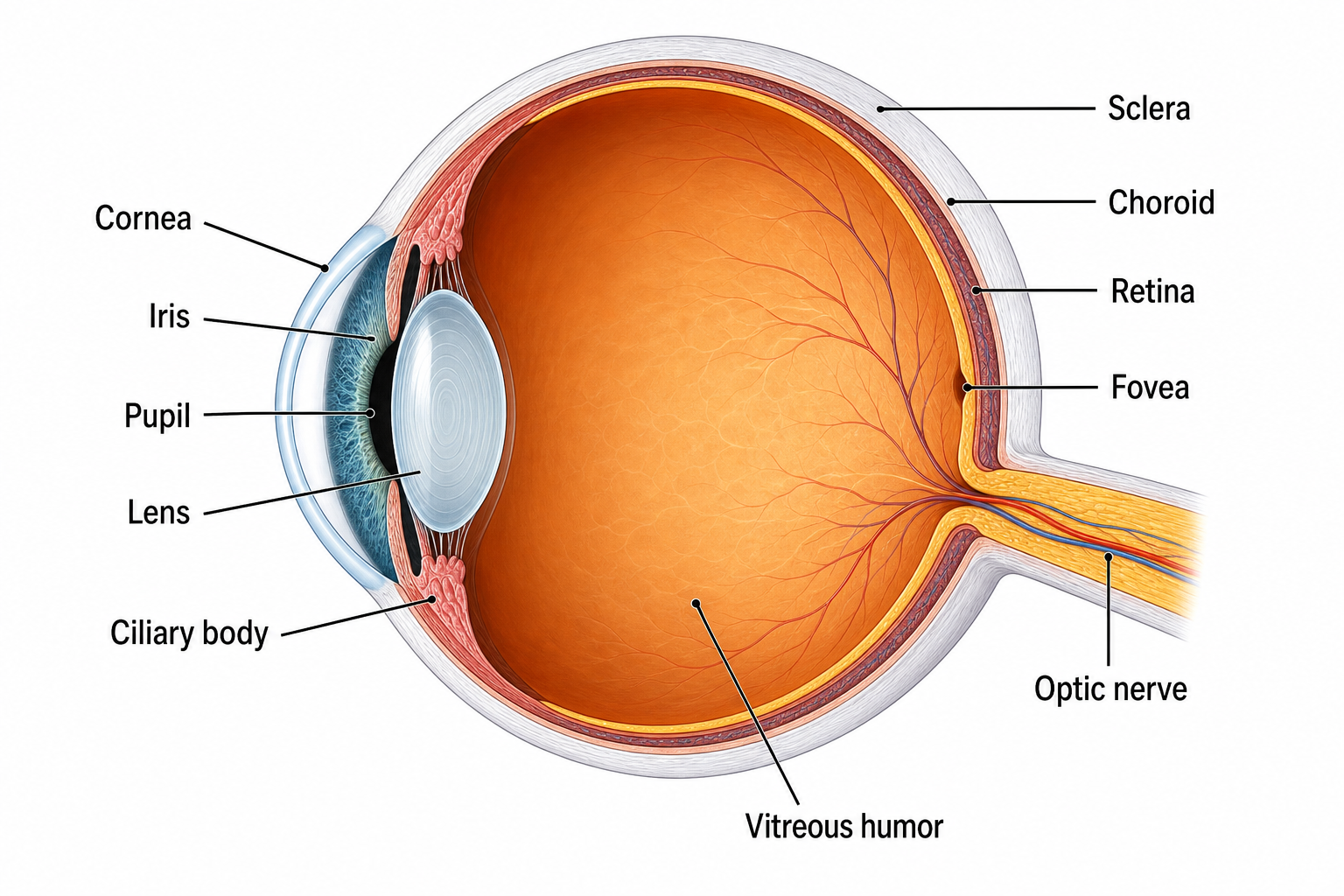
An eye anatomy diagram is a labeled illustration of the human eye that shows its structures and how they fit together. It usually presents a cross section of the eyeball so you can see external parts like the cornea and iris alongside internal parts like the lens, retina, and optic nerve. These diagrams turn a complex three-dimensional organ into a clear, readable picture, which is why they are a staple of biology, optometry, ophthalmology, and nursing study. SciDraw AI generates these diagrams from a short description, drawing tidy callout labels connected by leader lines so each part is easy to identify. The result is anatomically reasonable and clearly labeled, ready for slides, worksheets, and study notes.
Why use an eye anatomy diagram generator?
- Save hours of drawing time by generating a labeled diagram in seconds instead of sketching one by hand.
- Get clean, consistent labels so students can quickly learn the parts of the eye without clutter.
- Create custom views on demand, whether you need a cross section, a retina close-up, or the eye muscles.
- Produce high-resolution images that stay crisp on lecture slides, printed handouts, and exam materials.
- Adapt diagrams to your level, from a simple labeled overview to a more detailed structural breakdown.
How to make a labeled eye diagram
Start by typing a short description of the diagram you want, such as a labeled cross section of the human eye or a close-up of the retina. Name the specific structures you need labeled, like the cornea, iris, lens, retina, fovea, and optic nerve, and mention the view you prefer, such as a cross section or a front view. Choose the 4:3 aspect ratio for a balanced classroom layout and 2K resolution for sharp results. Click Generate Eye Diagram, then review the labeled image. If you want to adjust the level of detail or add or remove structures, refine your prompt and generate again. Before using a diagram for teaching or assessment, compare it against an authoritative anatomy reference to confirm every label is correct.
Parts of the eye
- Cornea: the clear front layer that protects the eye and bends incoming light to begin focusing.
- Iris and pupil: the colored ring that controls how much light enters, and the central opening it adjusts.
- Lens: the flexible structure behind the pupil that fine-tunes focus through accommodation.
- Retina: the light-sensitive inner layer, including the macula and fovea, that captures the image.
- Optic nerve: the bundle of fibers that carries visual signals from the retina to the brain.
- Sclera, choroid, and vitreous: the protective white wall, the nourishing middle layer, and the gel filling the eyeball.
Frequently asked questions
What are the parts of the eye?
The main parts of the eye include the cornea, iris, pupil, and lens at the front, the sclera and choroid forming the wall, and the retina, macula, fovea, and optic nerve at the back. The eyeball is filled with vitreous humor, and a watery aqueous humor sits in the front chambers. SciDraw AI can label all of these structures in a single diagram so each part is easy to find.
How do I make a labeled eye diagram online?
Describe the diagram you want in the generator, naming the view and the structures you need labeled, such as the cornea, iris, lens, retina, and optic nerve. Choose your aspect ratio and resolution, then click Generate Eye Diagram. SciDraw AI draws a clean diagram with tidy callout labels in seconds, and you can refine the prompt to add detail or adjust the view.
What types of eye diagrams can I create?
You can generate a fully labeled cross section, an eyeball cross section showing internal chambers, a retina close-up with the macula and fovea, a diagram of the six extraocular muscles, a lens accommodation comparison, and a front view of the external eye. Just describe the view you need and the structures you want labeled.
Are the diagrams accurate and clearly labeled?
SciDraw AI draws anatomically reasonable, clearly labeled diagrams with each structure connected to a tidy callout label, which makes them well suited for study and presentation. Because AI-generated images can occasionally place or label a structure imperfectly, you should always verify the diagram against an authoritative anatomy textbook or atlas before using it for teaching or assessment.
Is the eye anatomy diagram generator free to use?
You can start generating eye anatomy diagrams with SciDraw AI for free, and free credits refresh regularly so you can keep creating. If you need a higher volume of diagrams or more frequent generation, paid plans add more monthly credits. Each diagram uses a small number of credits per image.
Can I use these diagrams for biology or anatomy class?
Yes. The diagrams are designed for biology, optometry, ophthalmology, and nursing education, and work well in lecture slides, lab handouts, study guides, and worksheets. For classroom and exam use, confirm each label against a trusted reference so students learn the correct terminology.
Explore More Tools


Ready to draw the human eye?
Generate a clearly labeled eye anatomy diagram in seconds and bring your biology, optometry, or nursing lessons to life with SciDraw AI.