Zellmembran-Diagramm-Generator
Beschriftete, unbeschriftete und leere Zellmembran-Diagramme in Sekunden
Beschreibe den Membranaufbau, den du brauchst, und erhalte ein beschriftetes Referenzdiagramm, ein unbeschriftetes Diagramm zum Ausfüllen oder ein leeres Membran-Arbeitsblatt für den Biologieunterricht.

Beispiele für beschriftete und leere Zellmembran-Diagramme
Starte mit einem fertigen Beispiel oder beschreibe dein eigenes – vom vollständig beschrifteten Flüssig-Mosaik-Modell bis zum leeren Membran-Arbeitsblatt zum Ausfüllen.
Was macht dieser Zellmembran-Diagramm-Generator?
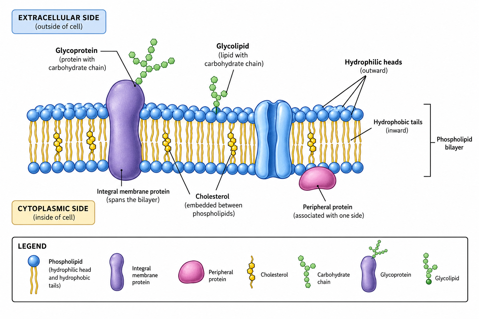
Er verwandelt die Beschreibung der Plasmamembran in ein klares, beschriftetes Diagramm nach dem Flüssig-Mosaik-Modell. Du beschreibst, welche Strukturen enthalten sein sollen – die Phospholipid-Doppelschicht, integrale und periphere Proteine, Kanal- und Carrier-Transportproteine, Cholesterin sowie Glykoproteine oder Glykolipide – und die KI zeichnet die Doppelschicht mit nach außen gerichteten hydrophilen Köpfen und nach innen weisenden hydrophoben Schwänzen, bettet die Proteine ein und fügt Textfelder und eine Legende hinzu, sodass die Struktur auf einen Blick klar ist – ganz ohne Illustrationssoftware oder manuelles Zeichnen. Aus derselben Beschreibung entsteht auch die Unterrichtsversion: Verlange eine unbeschriftete Membran mit leeren Bezugslinien, und du erhältst ein druckfertiges Arbeitsblatt zum Beschriften; erzeuge anschließend die vollständig beschriftete Fassung, und du hast das passende Lösungsblatt.
Warum einen Zellmembran-Diagramm-Generator nutzen
- Die Zellmembran ist ein zentrales Thema der Biologie, und ein klares Diagramm macht ihren Aufbau leicht verständlich.
- Eine korrekte Doppelschicht mit Proteinen, Cholesterin und Kohlenhydraten von Hand zu zeichnen kostet Zeit und Können.
- Beschriftete Bestandteile helfen Schülern und Lesern, jede Komponente mit ihrer Funktion zu verbinden.
- Lehrkräfte und Forschende brauchen klare, vorzeigbare Abbildungen für Unterricht, Arbeitsblätter, Veröffentlichungen und Folien.
- Aus einer Beschreibung neu zu erzeugen ist schneller, als die Membran jedes Mal neu zu zeichnen, wenn sich Beschriftungen oder Schwerpunkt ändern.
So erstellst du ein Zellmembran-Diagramm
Beschreibe die benötigte Membran und was hervorgehoben werden soll – zum Beispiel das vollständige Flüssig-Mosaik-Modell oder nur die Phospholipid-Doppelschicht. Gib an, welche Komponenten enthalten sein sollen: hydrophile Köpfe und hydrophobe Schwänze, integrale und periphere Proteine, Kanal- und Carrier-Proteine, Cholesterin sowie Glykoproteine oder Glykolipide. Notiere, ob Transportmechanismen gezeigt und die extrazelluläre und zytoplasmatische Seite markiert werden sollen. Erstelle die Abbildung, prüfe dann Ausrichtung, Komponenten und Beschriftungen und verfeinere sie, bis sie zu deinem Lehr- oder Berichtsstoff passt. Gib an, ob alle Bestandteile benannt werden sollen oder ob du eine unbeschriftete Fassung mit leeren Bezugslinien zum Ausfüllen brauchst. Für ein Arbeitsblatt hilft der Zusatz „weißer Hintergrund, druckfreundlich“.
Bestandteile eines Zellmembran-Diagramms
- Phospholipid-Doppelschicht – zwei Schichten mit hydrophilen Köpfen außen und hydrophoben Schwänzen innen.
- Integrale Proteine – Transmembranproteine, die in die Doppelschicht eingebettet sind, darunter Kanäle und Carrier.
- Periphere Proteine – Proteine, die an der inneren oder äußeren Membranoberfläche angeheftet sind.
- Cholesterin – Moleküle zwischen den Phospholipiden, die die Membranfluidität regulieren.
- Glykoproteine und Glykolipide – Kohlenhydratketten an der extrazellulären Oberfläche zur Erkennung.
- Transportwege – passiver (Diffusion, Osmose, erleichterte Diffusion) und aktiver Transport durch Proteine.
FAQ zum Zellmembran-Diagramm-Generator
Was ist das Flüssig-Mosaik-Modell?
Das Flüssig-Mosaik-Modell beschreibt die Zellmembran als flüssige Phospholipid-Doppelschicht mit einem Mosaik aus Proteinen, Cholesterin und Kohlenhydraten, die darin eingebettet oder daran angeheftet sind. Die Phospholipide und viele Proteine können sich seitlich bewegen, weshalb die Membran als flüssig bezeichnet wird.
Warum ist die Phospholipid-Doppelschicht mit Köpfen außen und Schwänzen innen angeordnet?
Jedes Phospholipid hat einen hydrophilen (wasserliebenden) Phosphatkopf und zwei hydrophobe (wasserabweisende) Fettsäureschwänze. Im Wasser weisen die Köpfe zur wässrigen extrazellulären Flüssigkeit und zum Zytoplasma, während sich die Schwänze nach innen vom Wasser weg ausrichten und so eine stabile Doppelschicht mit hydrophobem Kern bilden.
Was ist der Unterschied zwischen integralen und peripheren Proteinen?
Integrale Proteine sind in die Doppelschicht eingebettet und durchspannen sie oft vollständig als Transmembranproteine, darunter Kanal- und Carrier-Proteine. Periphere Proteine sind nicht im hydrophoben Kern eingebettet; sie heften sich an die innere oder äußere Membranoberfläche oder an integrale Proteine an.
Kann das Diagramm den Membrantransport zeigen?
Ja. Du kannst Transportmechanismen anfordern, etwa einfache Diffusion, Osmose, erleichterte Diffusion durch Kanal- und Carrier-Proteine (passiver Transport) sowie aktiven Transport über Pumpen, die ATP nutzen, mit Pfeilen, die die Bewegungsrichtung zeigen.
Welche Funktion hat Cholesterin in der Membran?
Cholesterin liegt zwischen den Phospholipiden und hilft, die Membranfluidität zu regulieren, sodass die Membran über einen Temperaturbereich hinweg stabil bleibt. Du kannst Cholesterinmoleküle in die Doppelschicht aufnehmen und im Diagramm beschriften.
Kann ich eine bearbeitbare Abbildung exportieren?
SciDraw AI kann in Vektorformate exportieren, sodass du Beschriftungen, Farben und Komponenten nachträglich anpassen kannst. Überprüfe das Diagramm immer anhand deines Lehrbuchs oder deiner Quelle, bevor du es verwendest.
Kann ich ein unbeschriftetes oder leeres Zellmembran-Diagramm erzeugen?
Ja. Beschreibe die Bestandteile, die gezeichnet werden sollen, und gib an, dass keine Beschriftungen gesetzt werden – oder dass nummerierte Bezugslinien auf leere Kästchen zeigen. Du bekommst dieselbe korrekte Phospholipid-Doppelschicht mit ihren Proteinen und dem Cholesterin, nur ohne jeden Text darauf, bereit zum Selbstbeschriften.
Kann ich das Zellmembran-Diagramm als Arbeitsblatt und Lösungsblatt verwenden?
Ja. Erzeuge zuerst die leere Doppelschicht mit nummerierten, unbeschrifteten Bezugslinien und drucke sie als Arbeitsblatt zum Beschriften. Erzeuge anschließend ein vollständig beschriftetes Flüssig-Mosaik-Modell als Lösungsblatt. Gib in beiden Prompts dieselbe Ansicht und dieselbe Nummerierung an, damit die beiden Blätter zusammenpassen.
Schreibst du an einem Paper, einer Abschlussarbeit oder einem Antrag?
SciDraw AI erstellt auch publikationsreife Forschungsabbildungen: Graphical Abstracts, Mechanismus-Abbildungen, Workflows und konzeptionelle Rahmenmodelle.
Weitere Tools entdecken
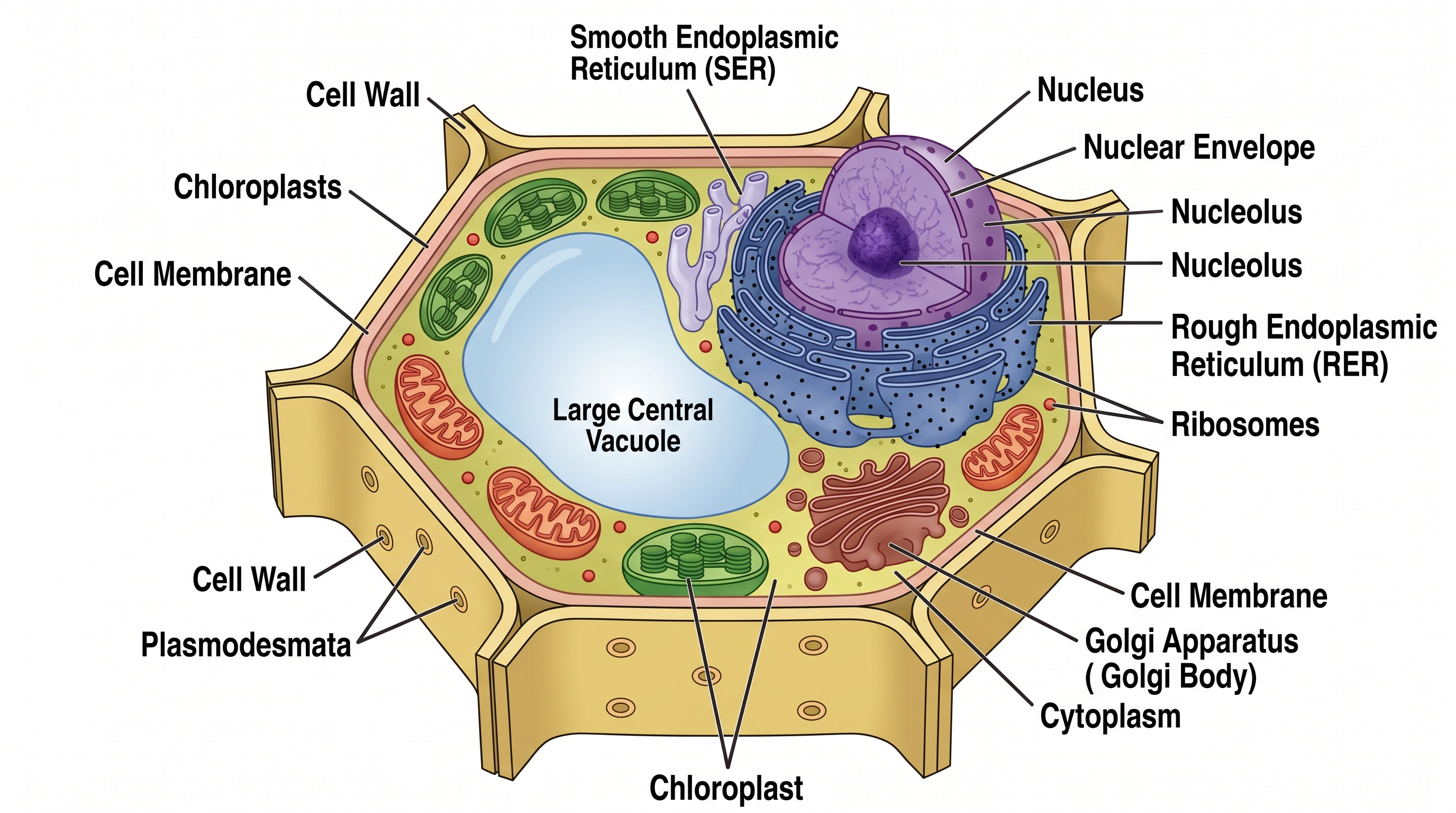
Pflanzenzell-Diagramm
Beschriftetes Pflanzenzell-Diagramm mit allen Organellen generieren.
Kostenlos testen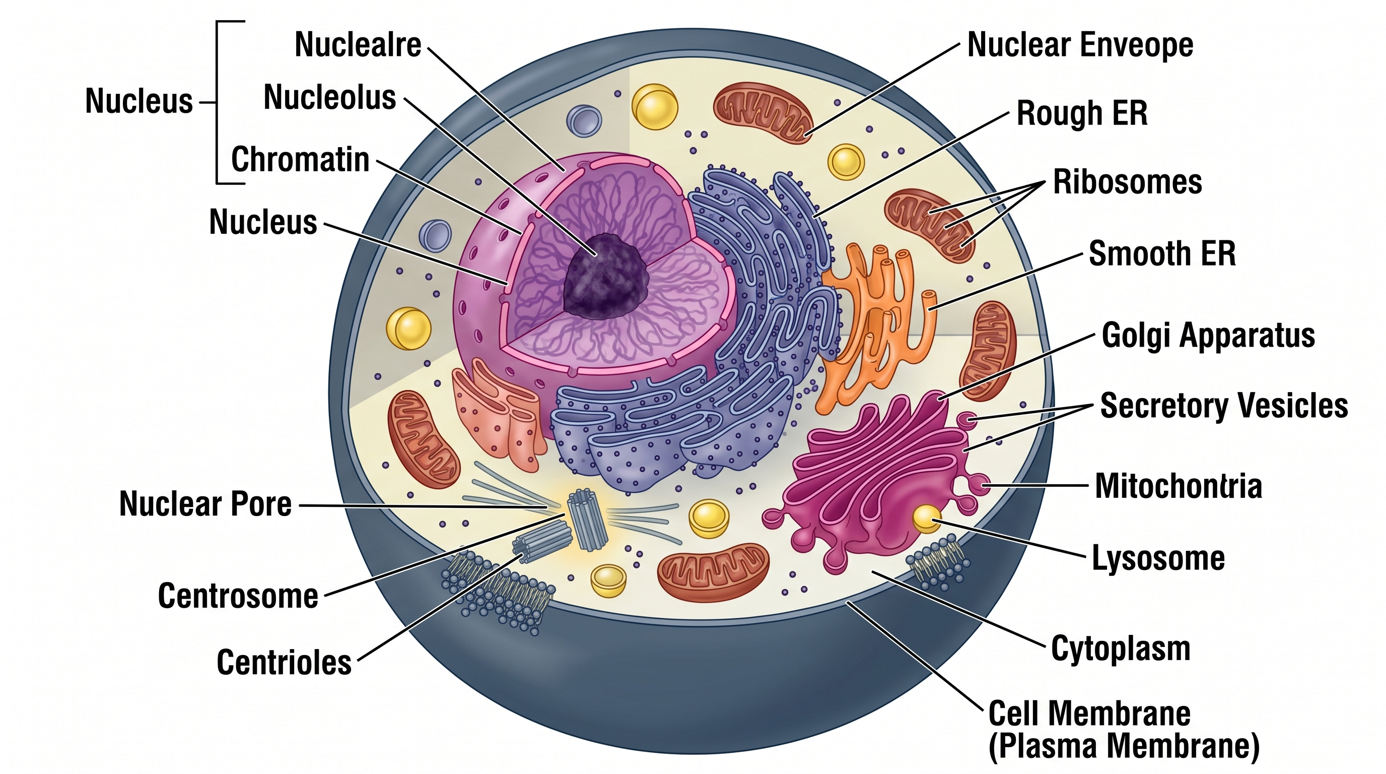
Tierzell-Diagramm
Beschriftetes Tierzell-Diagramm mit allen Organellen generieren.
Kostenlos testen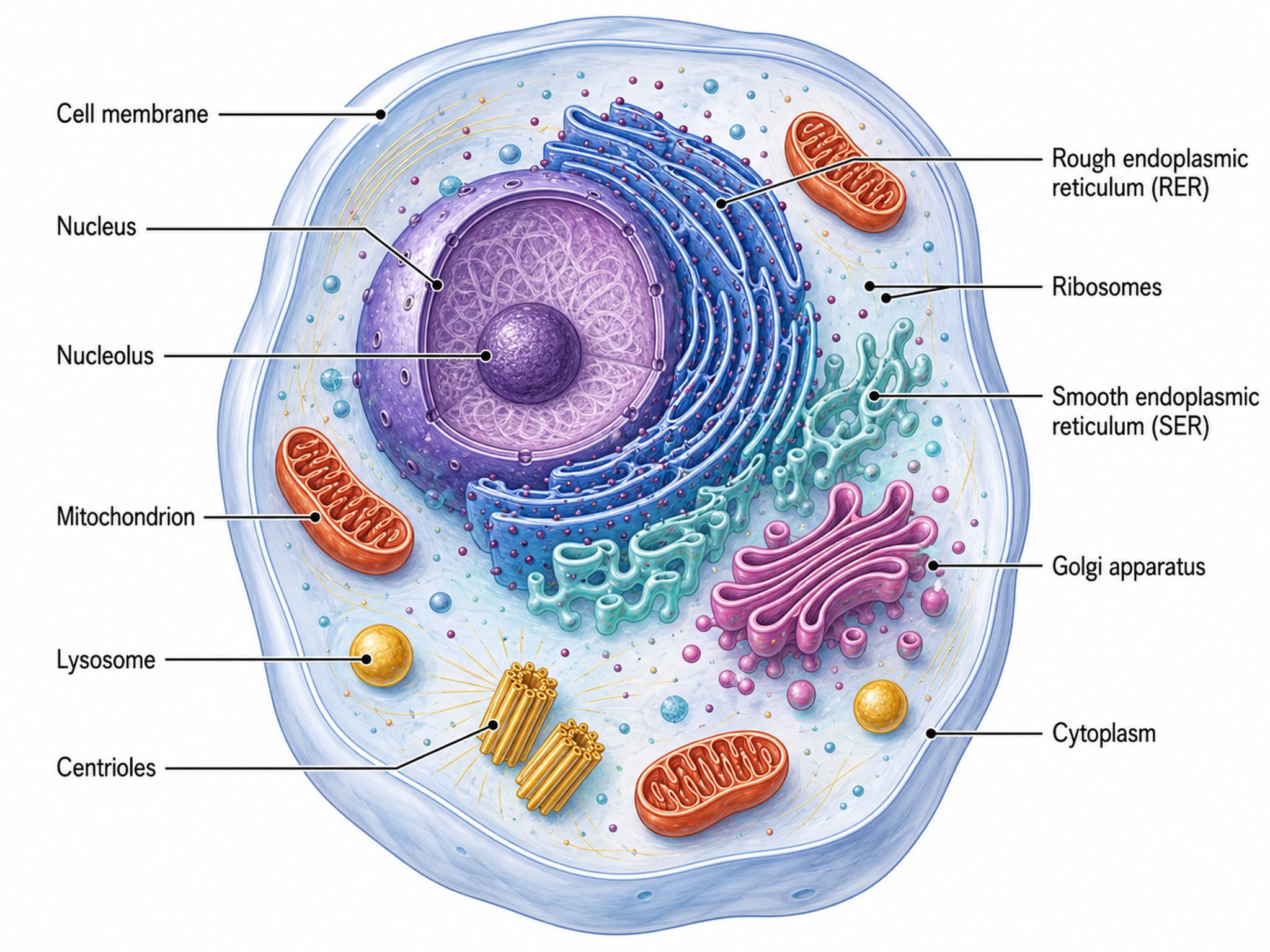
Zellillustrations-Generator
Zellbiologie-Diagramme und Organellen-Illustrationen erstellen
Kostenlos testenBrauchst du weitere Biologie-Abbildungen?
Erzeuge in Sekunden ein beschriftetes Zellmembran-Diagramm, eine unbeschriftete Fassung zum Beschriften oder ein leeres Membran-Arbeitsblatt. Perfekt für den Biologieunterricht.