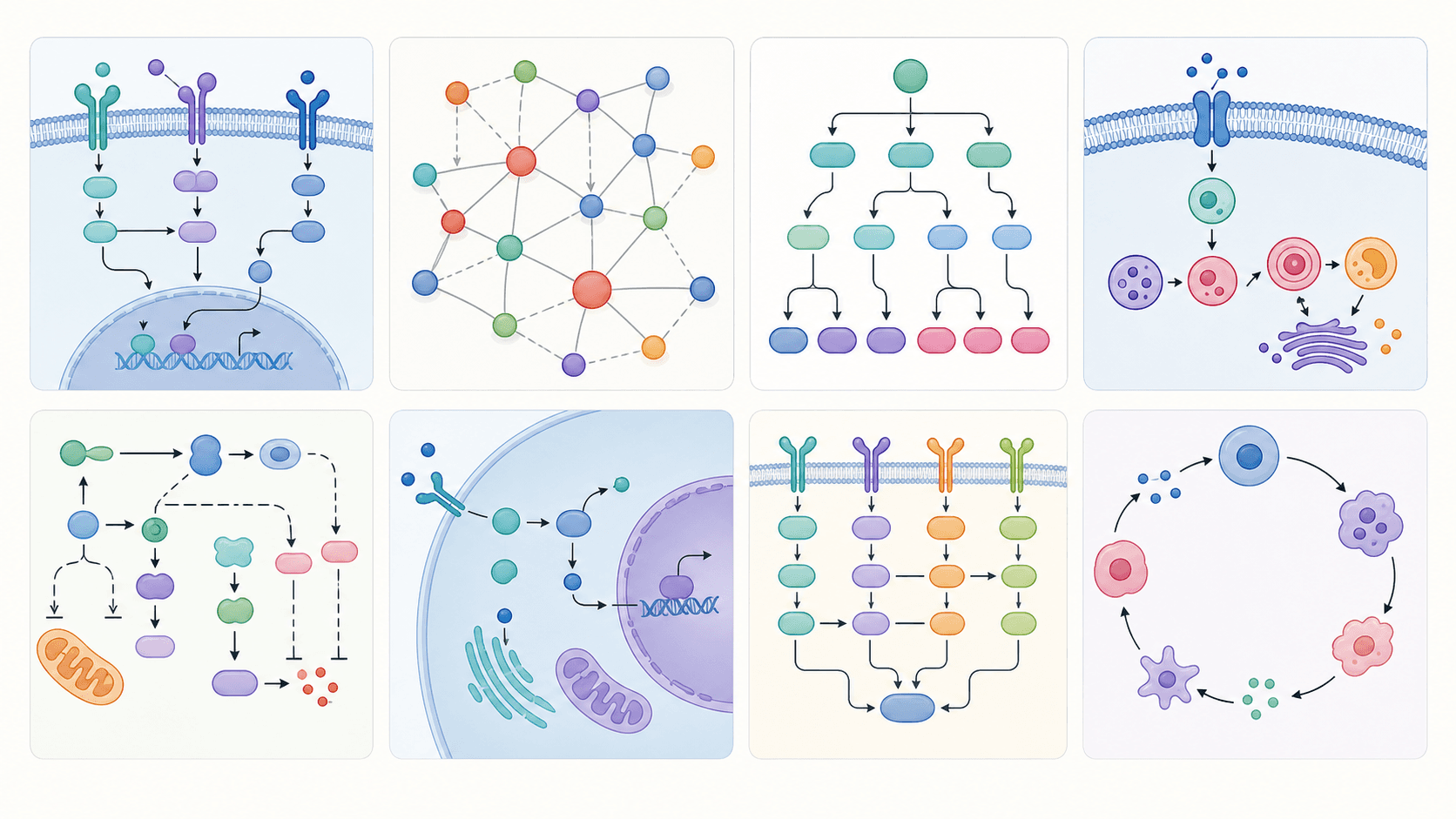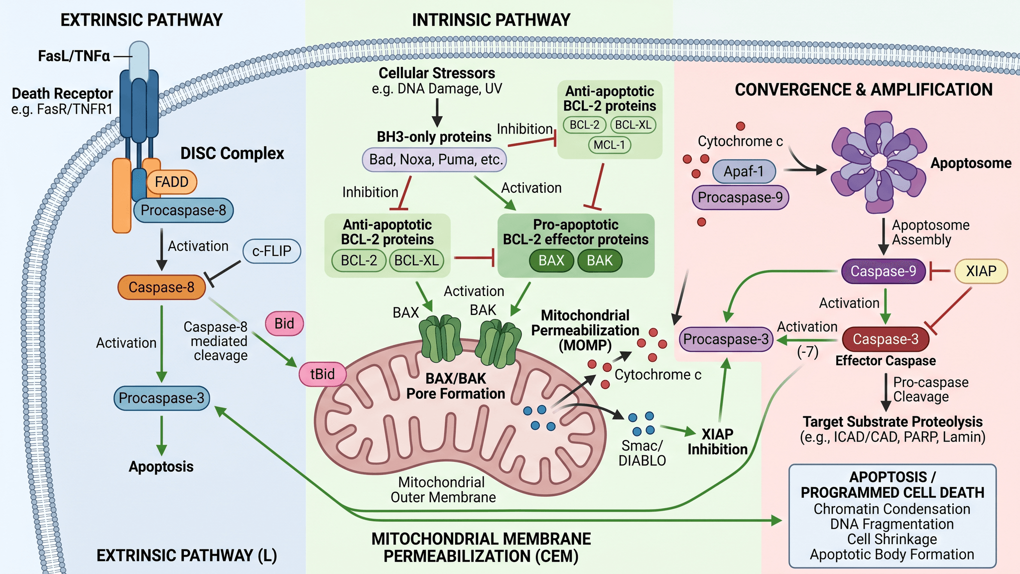
The apoptosis pathway is one of the most-drawn — and most-mislabeled — figures in cell biology. It packs two converging cascades (the intrinsic and extrinsic pathways), a dozen caspases and adaptors, the BCL-2 family balance, and a mitochondrial release step into a single frame. Drawn by hand, one wrong arrow turns activation into inhibition and changes the biology. This guide shows you how to generate a clean, accurate, publication-ready apoptosis pathway diagram with AI from a single sentence — then refine it by talking to the editor.
By the end of this guide you'll be able to:
- Generate a complete apoptosis pathway diagram — intrinsic and extrinsic — from one prompt.
- Label every key player correctly: death receptors, FADD, caspase-8, BAX/BAK, cytochrome c, Apaf-1, the apoptosome, caspase-9, and the executioner caspase-3.
- Show activation vs. inhibition so the BCL-2 family brake reads correctly.
- Adapt the prompt to your own study with a simple four-part template.
- Export an editable figure for your manuscript, slides, or poster.
Paste any prompt below into the Signaling Pathway Diagram Generator, then refine the result in the SciDraw AI editor — your image is the canvas and language is the tool.
A reusable prompt template for apoptosis figures
Weak results almost always come from vague prompts. Strong apoptosis diagram prompts have four parts:
- Subject — which pathway(s): intrinsic, extrinsic, or both converging.
- Steps and interactions — who activates or inhibits whom, in order.
- Labels — name every protein, complex, and organelle you want labeled.
- Style & layout — "flat vector, publication-ready, top-to-bottom flow, activation arrows and flat-end inhibition bars."
Template: "Draw the [intrinsic/extrinsic] apoptosis pathway showing [steps and interactions]. Label [proteins, complexes, organelles]. Use a clean flat-vector style with activation arrows and inhibition bars, top-to-bottom layout."
Copy-paste apoptosis pathway prompts
Intrinsic (mitochondrial) apoptosis pathway
The intrinsic pathway is triggered by internal stress — DNA damage, oxidative stress, growth-factor withdrawal — and runs through the mitochondrion.
- Draw the intrinsic (mitochondrial) apoptosis pathway: cellular stress activating pro-apoptotic BAX and BAK to form pores in the outer mitochondrial membrane, cytochrome c release into the cytosol, Apaf-1 binding to form the apoptosome, caspase-9 activation, and caspase-3 cleavage leading to apoptosis. Label BAX, BAK, BCL-2, cytochrome c, Apaf-1, apoptosome, caspase-9, caspase-3.
- Show the BCL-2 family balance: anti-apoptotic BCL-2 and BCL-xL inhibiting BAX/BAK (inhibition bars), and pro-apoptotic BH3-only proteins (BIM, BID, PUMA) tipping the balance toward pore formation.
Extrinsic (death receptor) apoptosis pathway
The extrinsic pathway starts at the cell surface when a death ligand binds its receptor.
- Draw the extrinsic apoptosis pathway: FasL binding the Fas (CD95) death receptor, recruitment of FADD, formation of the death-inducing signaling complex (DISC), caspase-8 activation, and direct cleavage of caspase-3. Label FasL, Fas/CD95, FADD, DISC, caspase-8, caspase-3.
- Illustrate TNF-α binding TNFR1, complex formation, and the branch point between survival (NF-κB) and apoptosis (caspase-8), as two diverging arrows.
Both pathways converging
- Draw both apoptosis pathways converging: the extrinsic pathway (death receptor → caspase-8) and the intrinsic pathway (mitochondrial stress → cytochrome c → apoptosome → caspase-9) both activating the executioner caspase-3, with caspase-8 cleaving BID to tBID as the cross-talk link. Two-column layout meeting at the bottom.
If your paper's figure is really about how a drug or stimulus triggers apoptosis — the pathway plus the treatment and the cellular outcome — try the mechanism figure generator.
How to get clean, accurate apoptosis diagrams
- Name every element you want labeled. The AI labels what you name — list "caspase-8, caspase-9, caspase-3, BAX, BAK, BCL-2, cytochrome c, Apaf-1."
- Specify direction explicitly. Use "activation arrow" and "flat-end inhibition bar" so the BCL-2 brake reads as inhibition, not activation.
- Choose a layout. Top-to-bottom suits a single cascade; a two-column converging layout suits both pathways at once.
- Mark the cross-talk. Tell the AI that caspase-8 cleaves BID to tBID, linking extrinsic to intrinsic — otherwise the pathways look disconnected.
- Iterate, don't restart. Refine with "recolor the mitochondrion" or "make caspase-3 the largest node" instead of rewriting the prompt.
Common mistakes (and how to fix them)
- Activation vs. inhibition unclear. The BCL-2 family is the classic trap. Fix: request "BCL-2 and BCL-xL shown with flat-end inhibition bars on BAX/BAK."
- Missing the apoptosome. Many drafts jump from cytochrome c straight to caspase-9. Fix: add "show Apaf-1 assembling into the apoptosome before caspase-9 activation."
- Caspase numbers swapped. Initiator vs. executioner caspases get confused. Fix: label them by role — "initiator caspase-8/9" and "executioner caspase-3."
- Overcrowded figure. Fix: ask to "simplify to the main steps" or split intrinsic and extrinsic into two panels.
- Garbled text (typical of generic image AI). Fix: SciDraw AI renders clean sans-serif labels — re-prompt the exact wording if needed.
Export and use your apoptosis figure
Once the pathway looks right, export it to editable SVG or PowerPoint (PPTX), or download a high-resolution image for your manuscript, slides, or poster. Want more pathway prompts to adapt? See our signaling pathway diagram prompts. Need to fix a label or translate it? See how to edit text and labels in an AI figure.
Frequently asked questions
What is the best AI tool to draw an apoptosis pathway diagram? SciDraw AI's Signaling Pathway Diagram Generator is built for publication-ready cell-death figures — intrinsic and extrinsic apoptosis, caspases, the BCL-2 family, and the apoptosome — with clean, accurate labels and editable export.
How do I show the difference between intrinsic and extrinsic apoptosis? Use a two-column layout: the extrinsic pathway (death receptor → caspase-8) on one side and the intrinsic pathway (mitochondrial stress → cytochrome c → apoptosome → caspase-9) on the other, both converging on caspase-3. Use prompt #5 above as a starting point.
Can I make an apoptosis diagram for free? Yes — you can start generating apoptosis pathway diagrams for free, then upgrade for more credits and editable SVG/PPTX export.
How do I label caspases correctly in my figure? Name them by role in the prompt — "initiator caspase-8 and caspase-9, executioner caspase-3" — and the AI will label each node. You can also rename any label afterward by telling the editor the exact change.
Is the apoptosis figure accurate enough for publication? It is designed for publication-ready output, but always review the biology for your specific system — especially activation vs. inhibition arrows — and correct any labels before submitting.
Start creating
Pick any prompt above, paste it into the Signaling Pathway Diagram Generator, and refine it in the SciDraw AI editor until it matches your study. From the BCL-2 balance to the executioner caspase, your next apoptosis figure is one sentence away.