
Radiology learners do not need random "pretty images". They need figures that match reading workflow: identify findings, localize anatomy, compare patterns, and support differential diagnosis.
This guide gives you a reusable prompt framework for CT, MRI, and chest X-ray (CXR) teaching visuals.
The Radiology Prompt Problem
Short prompts like "lung nodule CT" often fail because they omit:
- plane and view (axial/coronal/sagittal)
- lesion location and boundary behavior
- required labels and arrows
- comparison targets (benign vs malignant, disease A vs B)
- educational level (intern, resident, board prep)
Prompt Formula for Imaging Findings
Create a [CT/MRI/CXR] teaching illustration for [condition].
View: [axial/coronal/lateral/AP], with key anatomical landmarks.
Findings: [size, shape, density/signal, distribution, associated signs].
Layout: [single labeled image / side-by-side differential / progression timeline].
Labels: arrows + concise callouts + legend.
Style: clean medical textbook vector, white background, high readability.
24 High-Intent Prompt Templates
CT Templates
CT chest benign vs malignant lung nodule comparison, 2-panel layout.
Left: smooth margin, central calcification, stable size. Right: spiculated
margin, pleural retraction, interval growth. Add arrow callouts.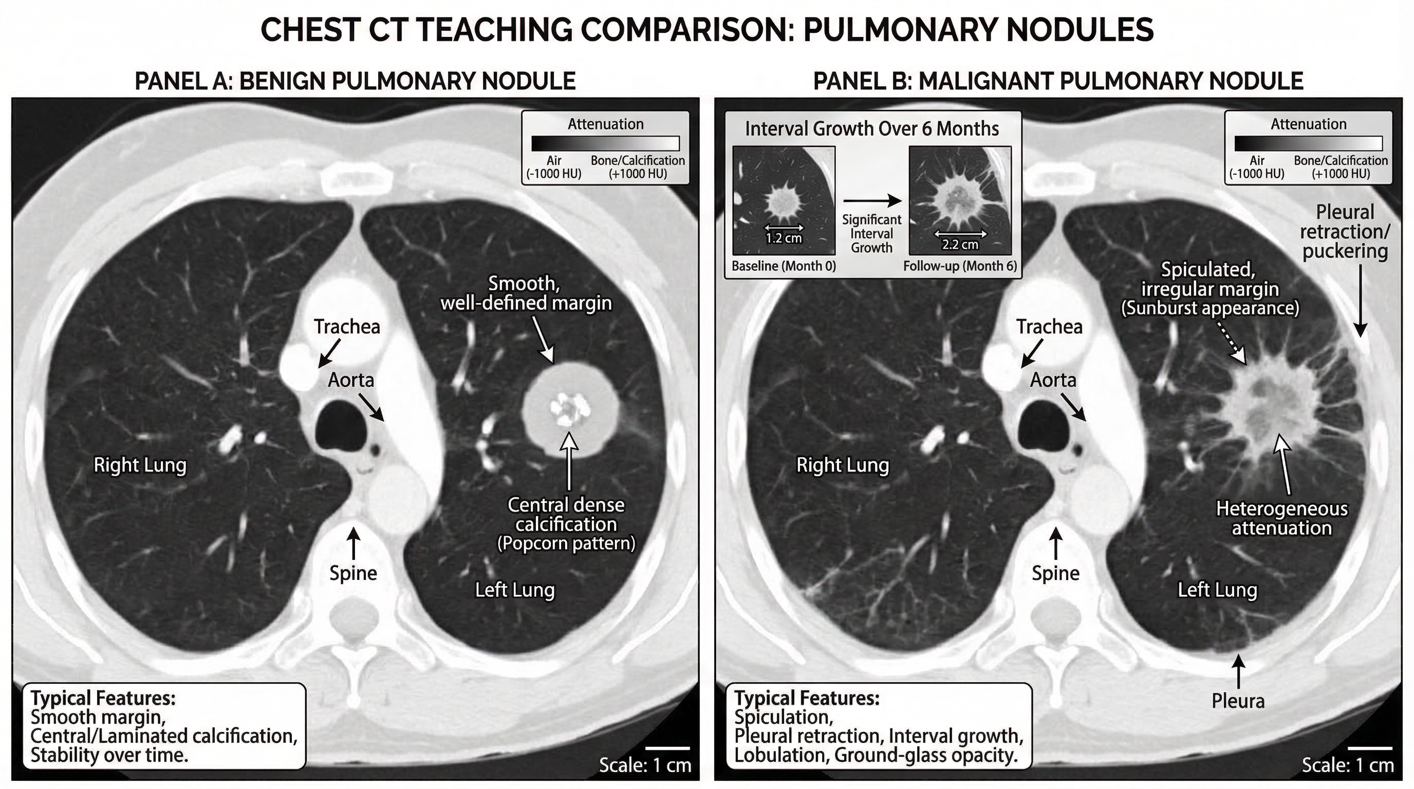
Acute appendicitis CT findings teaching figure, coronal and axial inset.
Label enlarged appendix diameter, wall thickening, periappendiceal fat stranding,
and appendicolith.
Pulmonary embolism CT angiography illustration with filling defect in segmental
artery, vessel course labeling, and right-heart strain indicators.MRI Templates
Acute ischemic stroke MRI sequence comparison.
DWI hyperintensity with ADC hypointensity, FLAIR mismatch concept, and lesion
territory map. Add timeline labels.
Multiple sclerosis brain MRI teaching panel with periventricular lesions,
Dawson fingers pattern, and sagittal FLAIR annotation.Pituitary adenoma MRI figure with sellar anatomy landmarks and contrast
enhancement differences vs normal pituitary tissue.CXR Templates
Pneumothorax CXR teaching figure with pleural line labeling,
absent peripheral lung markings, and tension signs.
Lobar pneumonia CXR map, right upper lobe consolidation with air bronchogram,
silhouette sign explanation, and differential notes.Pulmonary edema CXR pattern guide showing bat-wing opacities, Kerley B lines,
cardiomegaly, and small bilateral pleural effusion.Differential and Workflow Templates
CXR approach flowchart for dyspnea: airway, breathing, circulation,
lung fields, pleura, mediastinum, lines/tubes. Include quick red flags.
MRI ring-enhancing lesion differential chart: abscess, metastasis,
glioblastoma, toxoplasmosis. Add key discriminators in one panel.CT acute abdomen triage diagram comparing perforation, obstruction,
ischemia, and pancreatitis hallmark findings.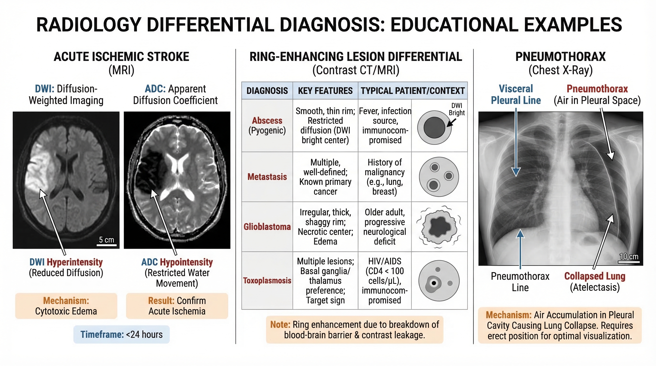
What Improves Clinical Usefulness
- always define imaging plane and modality
- include at least 3 discriminative findings
- use side-by-side comparison when teaching differential diagnosis
- keep labels short and diagnostic, not narrative
- design for slide readability first, journal polish second
Export-Ready Checklist
- modality and sequence are explicitly named
- anatomy landmarks are present
- lesion callouts are unambiguous
- differential cues are visible in one glance
- figure remains readable in presentation mode
Build faster, teach better, and standardize your imaging education assets.
Generate radiology teaching figures with SciDraw AI
Related Guides
- Medical Illustration Generator — create medical diagrams and teaching visuals
- Clinical Illustration Generator — create clinical and procedure-oriented figures
- Clinical Examination Diagram Prompts — step-by-step teaching figures
- ECG Diagram Prompts — 30 ECG teaching templates
- Biomedical Illustration Prompts — 50 AI prompts for biomedical visuals
- Book Illustration Maker Tool — create medical book illustrations


