
Clinical examination is procedural, but many teaching materials are still text heavy. Learners remember better when each step is visualized in sequence.
This article shows how to create exam-teaching diagrams for pulmonary, cardiovascular, abdominal, and neurologic examination using reusable prompt patterns.
Why Exam Diagrams Matter
Students often know the theory but miss sequence and positioning. A strong exam figure should answer:
- where to place hands/stethoscope
- what sequence to follow
- what positive findings look like
- how normal and abnormal differ
Prompt Formula for Examination Workflows
Create a clinical examination teaching diagram for [system].
Layout: [step-by-step vertical flow / 4-quadrant panel / checklist infographic].
Steps: inspection, palpation, percussion, auscultation (or system-specific order).
For each step, show hand/tool position + target area + key finding.
Add labels, arrows, and common mistakes in small callouts.
Style: clean medical education illustration, high clarity, white background.
20 Prompt Templates for Bedside Teaching
Pulmonary Examination
Pulmonary examination ladder technique diagram.
Show anterior and posterior chest auscultation points in sequence with numbered
markers, stethoscope positions, and side-by-side normal vs wheeze crackles notes.
Respiratory examination flowchart for OSCE:
inspection (work of breathing, cyanosis), palpation (trachea, expansion),
percussion zones, auscultation map. Include red-flag findings.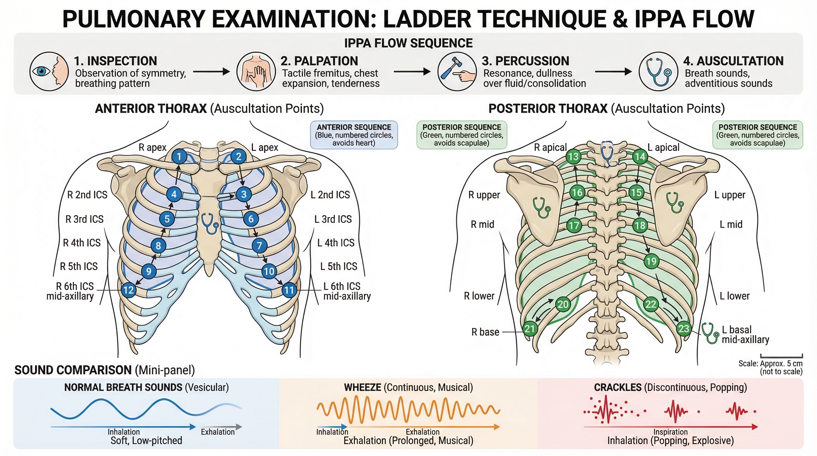
Cardiovascular Examination
Cardiac auscultation landmarks diagram with Aortic, Pulmonic, Tricuspid,
and Mitral areas. Add radiation directions and maneuvers (squat, Valsalva,
left lateral position).
Jugular venous pressure bedside assessment figure with patient angle,
landmark identification, and measurement method.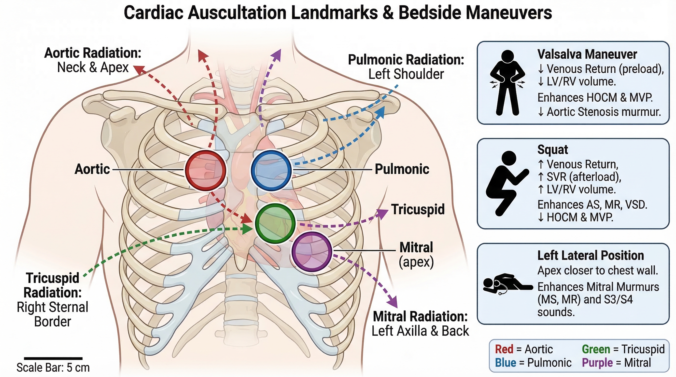
Abdominal Examination
Abdominal exam sequence infographic: inspection, auscultation, percussion,
palpation with quadrant order and pain-avoidance strategy.
Ascites examination panel comparing shifting dullness and fluid thrill methods,
with patient positioning and interpretation labels.Neurologic Examination
Cranial nerve examination workflow chart, CN I-XII grouped by practical station
sequence and key bedside maneuvers.
Weber and Rinne test teaching diagram with tuning fork placement, expected
normal pattern, and conductive vs sensorineural interpretation.Structured Assessment Templates
NIH Stroke Scale bedside overview diagram with section grouping, scoring logic,
and rapid interpretation summary.
General physical exam one-page checklist visual for ward rounds,
including vitals, HEENT, chest, heart, abdomen, neuro, extremities.Common Prompt Upgrades That Improve Output
- specify exact body orientation (sitting, supine, left lateral)
- request numbered step markers
- ask for "normal vs abnormal" in paired mini-panels
- include "common errors" callout boxes
- force concise labels under 6 words for projection clarity
Quick QA Before You Publish Slides
- Is sequence clinically correct?
- Are landmark positions unambiguous?
- Can students read labels from the back row?
- Does the figure include interpretation cues, not only anatomy?
- Is there one key takeaway per panel?
If your team teaches OSCE, bedside medicine, or clerkship bootcamps, these prompt patterns save time and improve consistency.
Build clinical teaching diagrams in SciDraw AI
Related Guides
- Medical Illustration Generator — create medical diagrams, anatomy figures, and teaching visuals
- Clinical Illustration Generator — create medical and clinical teaching illustrations with AI
- ECG Diagram Prompts — 30 templates for ECG teaching
- CT, MRI, and CXR Illustration Prompts — clinical imaging templates
- Biomedical Illustration Prompts — 50 AI prompts for biomedical visuals
- Medical Science Book Illustrations — create medical book visuals


")
")