LogoLogoSciDraw AI
  • Start Creating
  • Tools
  • Blog
  • Pricing
  • API
  • Education Discount
LogoLogoSciDraw AI

AI-powered scientific figure platform for researchers, students, educators, and science communicators. Create publication- and classroom-ready figures, graphical abstracts, TOC graphics, posters, and teaching illustrations in minutes. No design skills required.

EmailYouTubeXGitHubLinkedInInstagramStripe ClimateStripe Climate Contribution
Tools
  • AI Drawing
  • Scientific Data Visualization
  • Graphical Abstract Maker
  • Scientific Figure Maker
  • Image Converter
  • Vectorize Image
  • All Tools
Popular tools
  • Scientific Diagram Maker
  • Scientific Poster Maker
  • Research Poster Template
  • Plant Cell Diagram
  • Lewis Dot Structure Generator
  • Molecular Orbital Diagram Generator
  • PRISMA Flow Diagram Generator
  • Conceptual Framework Maker
Use Cases
  • For PhD Students
  • For Educators
  • For Journal Submission
  • BioRender Alternative
Resources
  • Blog
  • Scientific Chart Guide
  • Gallery
  • Published references
  • Media Kit
  • Developers
Company
  • About
  • Pricing
  • Affiliate
  • Institutional invoicing
  • Privacy Policy
  • Terms of Service
© 2026 SciDraw AI All Rights Reserved.
Figure Legend: Definition, Examples & How to Write One
2026/08/21

Figure Legend: Definition, Examples & How to Write One

What a figure legend is, with complete examples for microscopy, charts, blots, and schematics — plus a 4-part structure, common mistakes, and a submission checklist.

A figure legend (also called a figure caption) is the short block of text under a figure in a research paper that tells the reader what the figure shows and how to interpret it — the topic, what each panel contains, the key experimental context, and any abbreviations or statistics needed to read it correctly. A good legend lets a reader understand the figure without going back into the main text.

Here is what that looks like in practice:

Figure 2. Compound X reduces tumor growth in a dose-dependent manner. (A) Tumor volume in mice treated with vehicle, 10 mg/kg, or 50 mg/kg compound X for 21 days (n = 8 per group). (B) Representative tumor images at day 21. (C) Body weight over the treatment period. Data are mean ± SEM; *p < 0.05, **p < 0.01 vs. vehicle, one-way ANOVA with Tukey's post hoc test. Scale bar, 1 cm.

Every element has a job: a bolded title sentence states the finding, panels are walked in order, sample sizes and statistics are defined in place. The rest of this guide shows how to write legends like this — with more complete examples by figure type, the 4-part structure, and the mistakes reviewers flag most often.

You finished the figure at 11:40 p.m. The panels are aligned, the colors are finally consistent, and the scale bar is in the right corner. Then the real delay starts: the figure legend.

Most researchers do one of two things at this stage. They either write something too short and vague, like "Results of treatment group and control group," or they dump half of the Methods section under the figure and hope the reviewer sorts it out. Neither works well.

A strong figure legend does one practical job: it lets a reader understand the figure without constantly jumping back into the main text. That matters more than many authors realize, because figures and legends are often the first parts that reviewers read carefully.

This guide explains how to write a figure legend that is concise, informative, and publication-ready. It is written for research papers, not school lab reports or magazine captions, and it focuses on what actually helps a reader interpret the figure correctly.

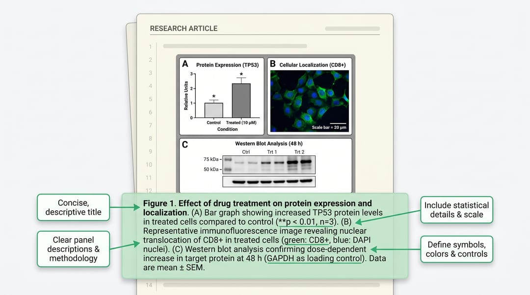
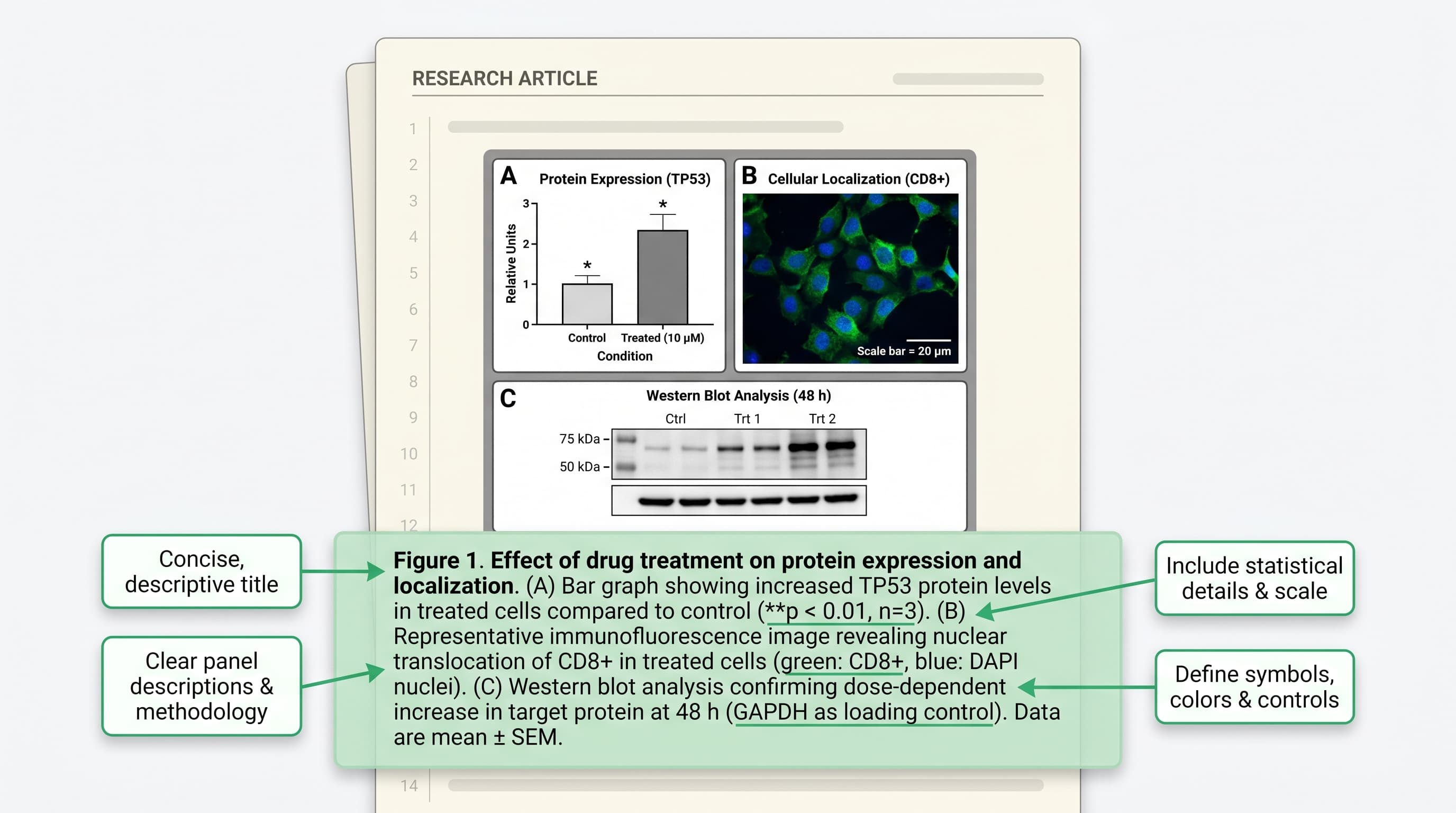
Editorial-style scientific cover showing manuscript page with figure panels and highlighted legend area A useful figure legend does not repeat the whole paper. It gives the reader exactly enough context to interpret the figure correctly.


What a figure legend is supposed to do

A figure legend is not just a label. It is also not a full narrative paragraph explaining the scientific significance of every result. Its job sits in between.

In most research papers, a good legend answers four questions:

  1. What is this figure about?
  2. What does each panel show?
  3. What details are necessary to interpret the visual correctly?
  4. What abbreviations, symbols, or statistics need to be defined here?

That means a figure legend usually needs to include:

  • the figure topic or main subject
  • the panel order, if the figure has panels such as A, B, and C
  • the experimental context necessary to read the figure
  • definitions of symbols, colors, abbreviations, and error bars
  • statistical details when the figure contains quantitative comparisons

What it usually does not need is a mini-discussion section. If the legend starts arguing for the significance of your finding instead of identifying what the figure contains, it has probably drifted too far.


The 5 most common figure legend mistakes

Before talking about structure, it is easier to see why many legends feel weak.

1. The legend is too generic

This is the classic:

Figure 2. Experimental results for the treatment and control groups.

That sentence tells the reader almost nothing. What results? Which treatment? What kind of readout? Microscopy? Bar chart? Flow cytometry? The legend is technically present, but functionally useless.

2. The legend repeats the Results section

The opposite problem is over-explaining:

Figure 2. Treatment with compound X significantly improved cell viability,
supporting our conclusion that the pathway is therapeutically actionable and
may represent a promising translational direction for future intervention.

That is interpretation, not figure guidance. The reader still does not know what each panel contains or how the data are displayed.

3. Panel order is unclear

If your figure has three or more panels, the legend should help the reader move through them in order. When that order is missing, the legend forces the reader to guess the logic of the layout.

4. Abbreviations are left undefined

Even if an abbreviation has already appeared in the main text, define it again in the legend when needed. Reviewers and readers often inspect figures independently.

5. Statistical details are omitted

If your bar chart includes error bars and significance markers, the reader needs to know what the error bars represent, what test was used, and how significance is encoded. Otherwise the figure is incomplete.


A simple structure that works for most figure legends

If you want a reusable formula, use this order:

  1. Start with a one-line topic statement.
  2. Describe panels in order.
  3. Add essential interpretation details.
  4. Define abbreviations and statistics.

That structure is flexible enough for most paper figures and rigid enough to keep you from wandering into filler.

Part 1: One-line topic statement

This should name the subject of the figure, not summarize the entire conclusion of the paper.

Good:

Figure 3. Effects of compound X on mitochondrial morphology and ATP production.

Weak:

Figure 3. Compound X improves mitochondrial function in an exciting and
clinically relevant way.

The first sentence helps the reader orient. The second sentence sounds like a conclusion slide.

Part 2: Panel-by-panel description

When panels are labeled, describe them in order.

For example:

(A) Representative fluorescence images of control and compound X-treated cells.
(B) Quantification of mitochondrial fragmentation.
(C) Intracellular ATP levels measured after 24 h treatment.

This is where many authors either become too vague or too wordy. The best version is usually concrete and compact.

Part 3: Essential interpretation details

This includes information that the reader needs in order to interpret the figure correctly, such as:

  • treatment duration
  • species, cell type, or sample type
  • stain or marker name
  • scale bar information
  • meaning of colors or symbols

Not every figure needs every item. The key question is simple: if this detail disappeared, would the figure become ambiguous?

Part 4: Abbreviations and statistics

For quantitative figures, do not leave the technical reading rules unstated.

Typical items include:

  • what error bars represent
  • what n means
  • which statistical test was used
  • p-value notation
  • abbreviation definitions

Example:

Data are shown as mean ± SD (n = 3 independent experiments). Statistical
significance was assessed by one-way ANOVA with Tukey's multiple-comparison
test. ns, not significant; *P < 0.05; **P < 0.01.

A worked example: weak legend vs strong legend

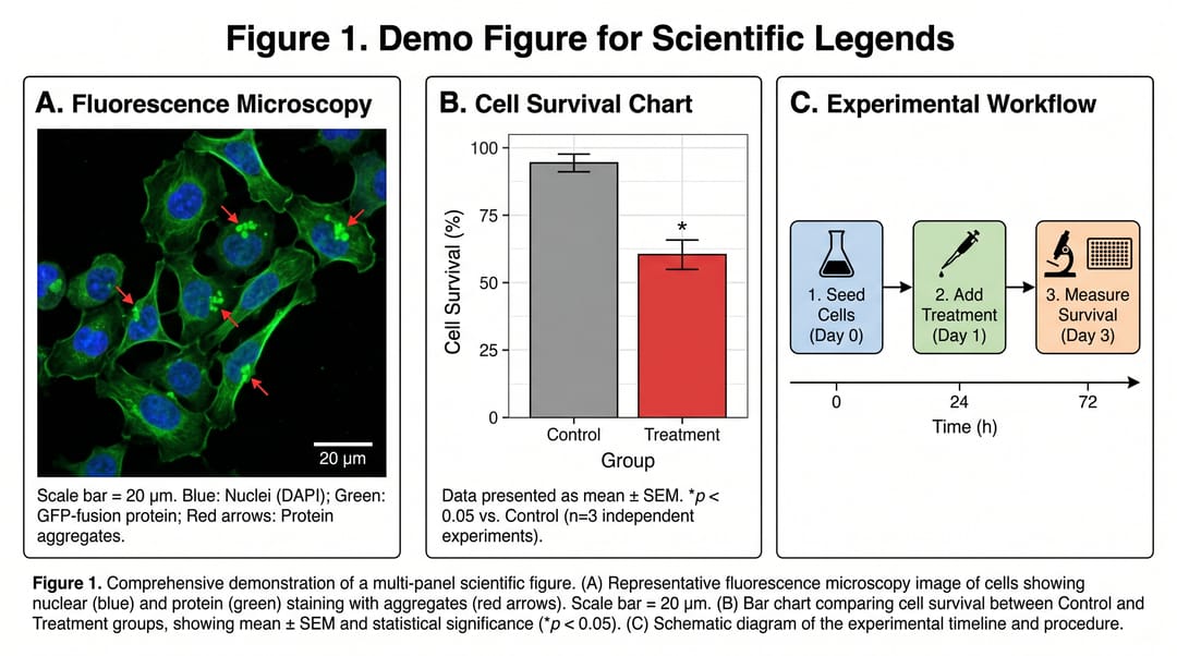
Below is a mock multi-panel figure used only to show how legend writing changes readability.

Mock multi-panel scientific figure with microscopy, bar chart, and workflow panels labeled A-C A demo figure with microscopy, quantification, and workflow panels. It is useful because it includes the kinds of mixed visual elements that often produce weak legends.

Weak version

Figure 1. Results of the experiment.
(A) Cells.
(B) Quantification results.
(C) Workflow of the study.

This is short, but it is not efficient. The reader still has to infer:

  • what kind of cells these are
  • what was being compared
  • what was quantified
  • whether the workflow shows treatment timing, sample processing, or analysis

Stronger version

Figure 1. Experimental overview and representative readouts of the treatment
effect. (A) Representative fluorescence microscopy images of control and
treated cells, showing mitochondrial signal after 24 h exposure; scale bar,
20 µm. (B) Quantification of normalized signal intensity in control and treated
groups. Bars represent mean ± SD from three independent experiments.
(C) Schematic workflow of treatment, imaging, and downstream quantification.
*P < 0.05 by unpaired two-tailed t test.

That version is still concise, but it does much more work. It tells the reader:

  • what the figure is about
  • how to read the panel sequence
  • what kind of image is shown
  • what the quantification represents
  • how the statistics should be interpreted

It is not perfect for every journal, but it is already far closer to a submission-ready legend than the weak version.


Complete figure legend examples by figure type

The fastest way to internalize the structure is to see finished legends. Each example below is publication-style and ready to adapt — swap in your own conditions, sample sizes, and statistics.

Fluorescence microscopy:

Figure 3. NLRP3 localizes to mitochondria upon inflammasome activation. Confocal images of THP-1 macrophages stained for NLRP3 (green), TOM20 (red), and DAPI (blue) after treatment with LPS + nigericin or vehicle for 6 h. Right panels show enlargements of the boxed regions. Images are representative of three independent experiments. Scale bars, 10 μm (main), 2 μm (inset).

Bar chart with statistics:

Figure 1. Drought stress reduces photosynthetic rate across all three cultivars. Net CO₂ assimilation rate in cultivars A, B, and C under well-watered and drought conditions (n = 6 plants per group, two growing seasons pooled). Bars show mean ± SD; individual points are single plants. **p < 0.01, ns = not significant, two-way ANOVA with Šídák's multiple comparisons test.

Western blot:

Figure 4. Compound Y inhibits ERK phosphorylation without affecting total ERK. (A) Immunoblots of p-ERK1/2, total ERK1/2, and β-actin in HeLa cells treated with the indicated concentrations of compound Y for 30 min. (B) Quantification of p-ERK/ERK ratio normalized to untreated control (n = 3 biological replicates). Data are mean ± SEM; *p < 0.05, one-sample t test vs. 1.0.

Workflow / schematic:

Figure 1. Study design and participant flow. Adults with type 2 diabetes (N = 248) were randomized 1:1 to the intervention or usual-care arm. Assessments occurred at baseline, 12 weeks, and 24 weeks. Created with SciDraw AI. Abbreviations: HbA1c, glycated hemoglobin; ITT, intention-to-treat.

Survival analysis:

Figure 5. High TREM2 expression is associated with shorter overall survival. Kaplan–Meier curves for patients stratified by median TREM2 expression (high, n = 187; low, n = 186; TCGA-LUAD cohort). Tick marks indicate censored patients. p value from two-sided log-rank test; HR, hazard ratio with 95% CI from univariable Cox regression.

If you already have the figure and want a first draft in this format, the AI Figure Legend Generator produces a structured legend from your figure and methods notes that you can then edit down.

What to include for different figure types

Different figures need different emphasis. The general structure stays the same, but the details shift.

Microscopy figures

Usually include:

  • specimen or cell type
  • stain, marker, or imaging channel
  • treatment or condition
  • scale bar
  • what is highlighted, if arrows or outlines are used

Common omission: authors mention the image but forget to define what the colors or markers mean.

Bar charts and quantitative panels

Usually include:

  • what is being measured
  • what the groups are
  • what the error bars represent
  • sample size or number of replicates
  • statistical test and significance notation

Common omission: authors show significance asterisks but never define them.

Workflow or schematic figures

Usually include:

  • what the workflow represents
  • step order
  • major conditions or sample categories
  • meaning of color coding or symbols

Common omission: the legend names the workflow but never explains what the color blocks mean.

Multi-panel mixed figures

These are the most common in research papers and also the easiest to write badly. The fix is simple: resist the urge to describe the figure globally in one long paragraph. Panel-by-panel structure is usually more readable.


Figure legend vs caption: are they the same?

In many journals, the terms are used loosely, and authors often mean roughly the same thing when they say "figure caption" or "figure legend." In practice, though, most researchers use figure legend to mean the full explanatory text that sits below a scientific figure.

That distinction matters because many authors write a legend as if it were only a short caption line. For research papers, that is usually too thin. A scientific figure legend often has to carry abbreviations, conditions, and statistics that a magazine-style caption would never include.

So if you are writing for journal submission, the safer assumption is:

  • a caption can be short
  • a figure legend usually needs to be self-contained

A fast checklist before submission

Before finalizing a legend, run through this checklist:

  • Can a reader understand what the figure is about from the first sentence?
  • If there are panels, are they described in order?
  • Are all abbreviations defined where needed?
  • Are colors, symbols, arrows, and labels explained if they are not obvious?
  • Are scale bars identified for image-based panels?
  • Are error bars defined for quantitative panels?
  • Is n stated clearly?
  • Is the statistical test named if significance is shown?
  • Does the legend avoid over-interpreting the result?
  • Could this figure be understood without rereading the whole Results section?

If you answer "no" to two or more of those questions, the legend probably still needs work.


A practical writing habit that saves time

The easiest way to write better figure legends is not to wait until the very end of the manuscript.

Instead, write a rough legend as soon as the figure layout is stable. At that point, you still remember:

  • why each panel exists
  • what each color means
  • which comparisons matter
  • what details are likely to confuse a reader

When authors postpone legends until submission week, they usually end up reconstructing their own reasoning from memory. That is why late-stage legends often sound generic.


Final takeaway

A good figure legend is not decorative. It is part of the figure itself.

If the figure carries the visual logic of your result, the legend carries the reading instructions. It tells the reader what they are looking at, how to move through it, and what technical details matter for interpretation. When that guidance is missing, even a good figure becomes slower to understand.

If you already have a figure and want a fast first draft of the legend, try the SciDraw AI Figure Legend tool. It will not replace final scientific judgment, but it can give you a much stronger starting point than a blank text box.


Related guides

  • How to Draw Scientific Figures: The Complete 2026 Guide
  • Nature, Science & Cell Figure Size, DPI & Format Requirements
  • Graphical Abstract Examples by Field
All Posts

Author

avatar for Davie Chen / SciDraw AI
Davie Chen / SciDraw AI

Researcher

Davie Chen is a researcher at the Faculty of Animation and Intermedia, University of Arts in Poznan, studying generative AI for scientific figure creation, patent illustration, and manuscript drafting. SciDraw AI is one of the research-to-product tools built from this work.

Author profile

Categories

What a figure legend is supposed to doThe 5 most common figure legend mistakes1. The legend is too generic2. The legend repeats the Results section3. Panel order is unclear4. Abbreviations are left undefined5. Statistical details are omittedA simple structure that works for most figure legendsPart 1: One-line topic statementPart 2: Panel-by-panel descriptionPart 3: Essential interpretation detailsPart 4: Abbreviations and statisticsA worked example: weak legend vs strong legendWeak versionStronger versionComplete figure legend examples by figure typeWhat to include for different figure typesMicroscopy figuresBar charts and quantitative panelsWorkflow or schematic figuresMulti-panel mixed figuresFigure legend vs caption: are they the same?A fast checklist before submission

More Posts

Create your scientific figures with AI

Thousands of researchers use SciDraw AI to make publication-ready figures for papers, grants, and journal submissions — in minutes, with no design skills.

Start for free
A practical writing habit that saves time
Final takeaway
Related guides
Tutorials
How to Add an Element, Label, or Arrow to a Figure with AI (2026)
Tutorials

How to Add an Element, Label, or Arrow to a Figure with AI (2026)

Add an element, label, or arrow to a scientific figure without redrawing it — drop in a new organelle, annotation, leader line, or directional arrow just by telling the AI what to add and where. Copy-paste prompts, a reusable template, and an AI figure editor that does the work.

avatar for Davie Chen / SciDraw AI
Davie Chen / SciDraw AI
2026/06/18
How to Draw a Scientific Figure for Your Paper: A 5-Step AI Workflow from Abstract to Submission
Tutorials

How to Draw a Scientific Figure for Your Paper: A 5-Step AI Workflow from Abstract to Submission

A reusable 5-step workflow for paper figures: define the central message, split into panels, write a structured prompt, then check against submission standards. Includes scientific-figure prompt templates and FAQ.

avatar for Davie Chen / SciDraw AI
Davie Chen / SciDraw AI
2026/06/18
How to Draw Biomedical Paper Figures with AI: A Unified Workflow for Cell, Pathway, and Workflow Diagrams
Tutorials

How to Draw Biomedical Paper Figures with AI: A Unified Workflow for Cell, Pathway, and Workflow Diagrams

How to keep cell diagrams, signaling pathway figures, and experimental workflows visually consistent across one biomedical paper — split figures into a background layer, mechanism layer, and experiment layer, then generate editable drafts with structured prompts. Includes prompt templates.

avatar for Davie Chen / SciDraw AI
Davie Chen / SciDraw AI
2026/06/18
Explore Our Tools
Figure Legend·Scientific Figure Maker·Graphical Abstract Maker·AI Scientific Illustration