
細胞生物学は、科学研究の中で最もイラストを多用する分野です。ここ数ヶ月だけでも、SciDraw AIでは1,300以上の細胞生物学および生物医学のイラストが作成されており、世界中の研究者がシグナル伝達経路、分子メカニズム、実験ワークフローを可視化するためのより優れたツールを必要としていることは明らかです。
白色脂肪細胞のベージュ化(browning)経路からCRISPR遺伝子編集のメカニズム、薬物動態から動物実験プロトコルまで、これらは論文の採否を左右する重要な図表となります。本ガイドでは、研究者コミュニティの実際の事例を用いて、それぞれのタイプの作成方法を紹介します。
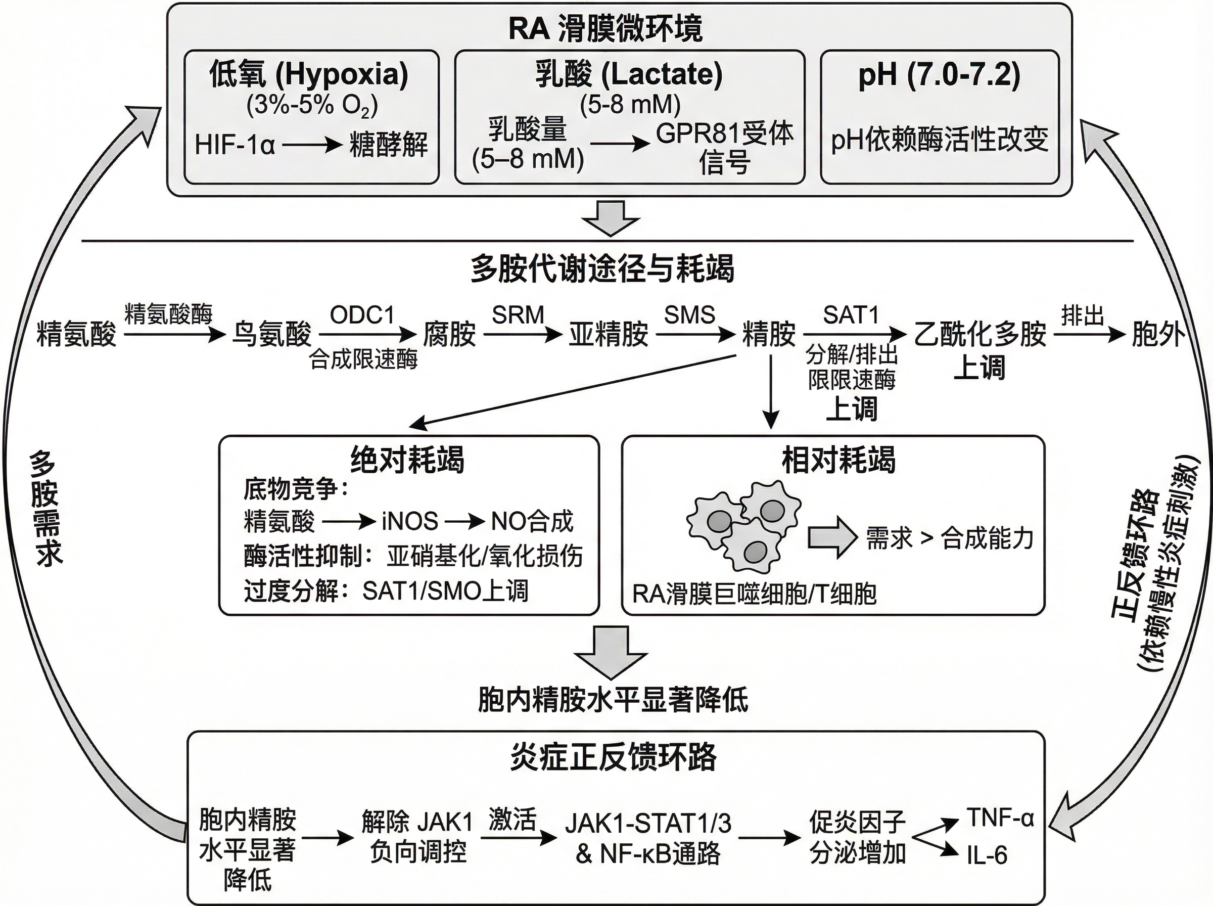 代謝ストレス経路を示す関節リウマチ滑膜微小環境のAI生成イラスト
研究者が実際に描いているもの
ユーザーコミュニティからの数千の実際のプロンプトに基づくと、最も人気のある細胞生物学イラストのタイプは以下の通りです。
- 細胞シグナル伝達経路(pathway, signaling, activation — 全生物医学プロンプトの10%以上で使用)
- 分子メカニズム(protein, molecular, mechanism — 最も多いキーワード)
- 薬物作用と薬理学(treatment, clinical, effects)
- 動物実験の模式図(mouse gavage, injection protocols)
- 遺伝子発現と調節(expression, transcription, CRISPR)
シグナル伝達経路のイラスト
シグナル伝達経路は、生物医学イラストの中で最もリクエストが多いタイプです。研究者は、シグナルが細胞膜受容体からキナーゼの連鎖を経て核内の転写因子へとどのように伝わるかを示す必要があります。
脂肪細胞のベージュ化経路
あるユーザーは、脂肪細胞のベージュ化に対する中医学的介入に関する、非常に説得力のあるイラストを作成しました。
Schematic diagram of the scientific mechanism, cell biology style,
browning pathway of white adipocytes.
On the left: Traditional Chinese medicine intervention
labeled "Spleen-tonifying and Turbidity-dissolving Formula."
Center: White adipocyte transitioning to beige adipocyte,
showing UCP1 upregulation, mitochondrial biogenesis,
β3-adrenergic receptor activation.
Arrows indicating AMPK/PGC-1α signaling cascade.
Clean white background, BioRender-style scientific illustration.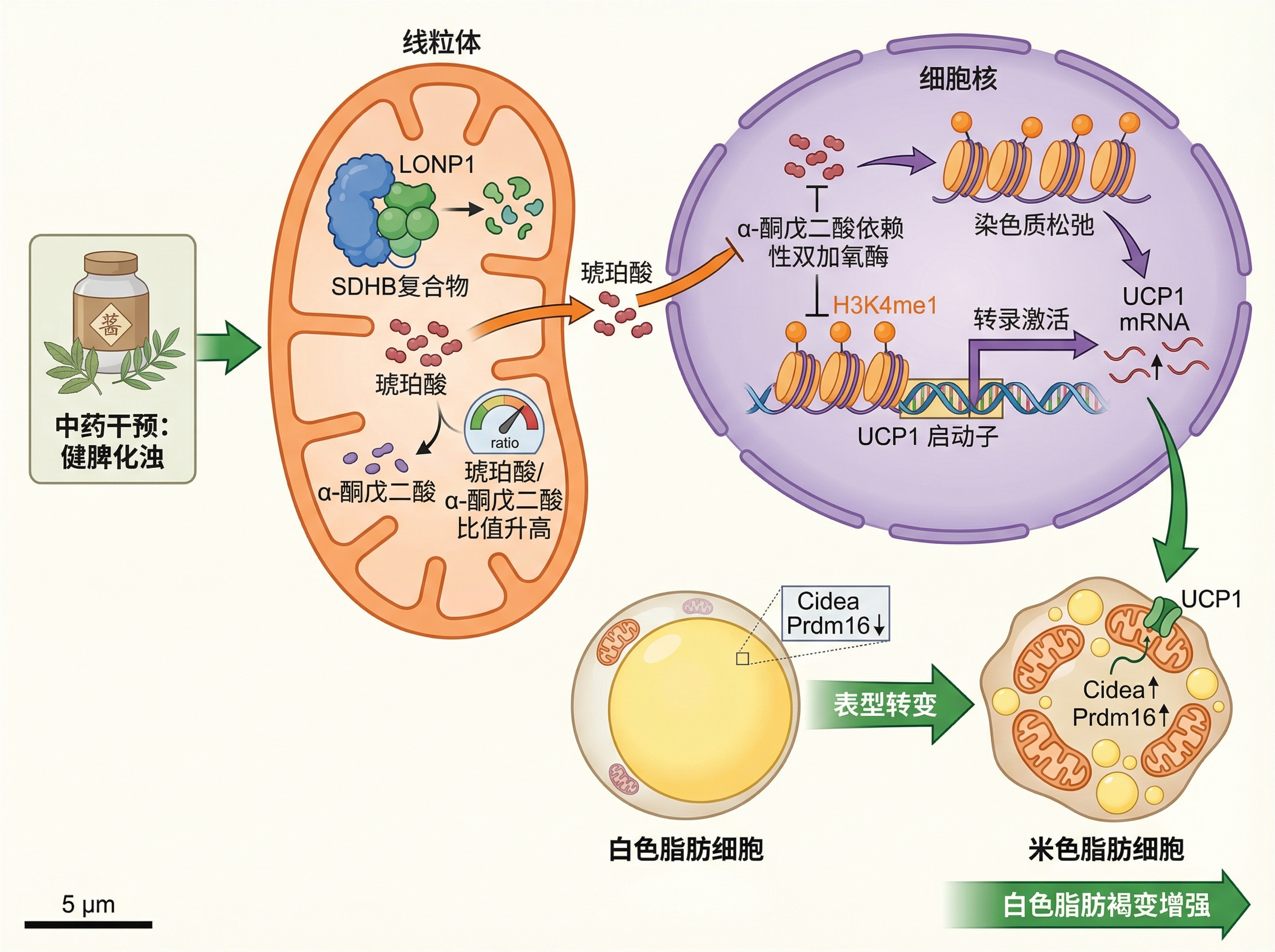 中医学的介入を伴う白色脂肪細胞のベージュ化経路
免疫回避とウイルスの経路
ウイルスのメカニズムのイラストは、免疫学やウイルス学の論文において不可欠です。
Porcine reproductive and respiratory syndrome virus (PRRSV)
immune evasion mechanism illustration.
Show rapid genetic mutation rate and host immune system evasion,
vaccine efficacy limitations due to viral diversity.
Include viral entry, replication cycle, and immune checkpoint modulation.
Schematic diagram style suitable for veterinary science journal.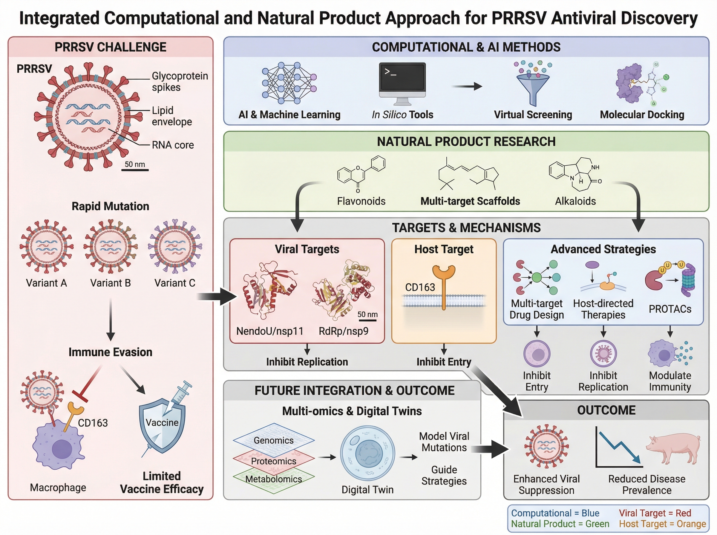 PRRSVの免疫回避メカニズムとワクチン設計の課題
分子メカニズム図
「メカニズム(Mechanism)」という言葉は、生物医学プロンプト全体の約16%に登場します。研究者は、分子レベルでの因果関係を示す必要があります。
薬物誘発性代謝経路
Valproic acid (VPA)-induced hyperammonemic encephalopathy (VHE)
mechanism illustration.
Show urea cycle impairment and mitochondrial dysfunction.
VPA reduces N-acetylglutamate levels affecting CPS1 activity.
Include: hepatocyte mitochondria, urea cycle enzymes,
ammonia accumulation pathway, blood-brain barrier crossing.
Schematic diagram suitable for pharmacology journal publication.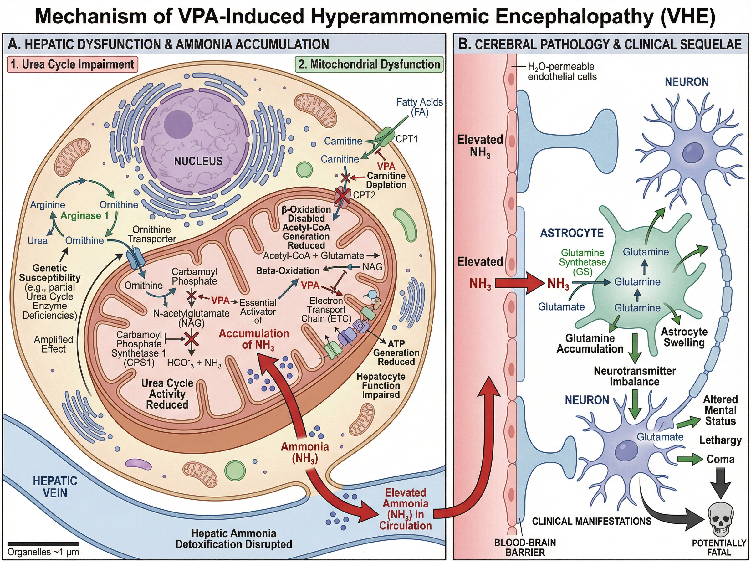 バルプロ酸誘発性高アンモニア血症性脳症のメカニズム
DNA損傷と修復経路
Model diagram for project proposal background section.
Radiation-induced DNA damage types (SSB, DSB, base damage),
DNA repair pathways including:
- Base excision repair (BER)
- Nucleotide excision repair (NER)
- Homologous recombination (HR)
- Non-homologous end joining (NHEJ)
Show pathway selection decision points and key proteins.
Academic paper style, suitable for radiobiology journal.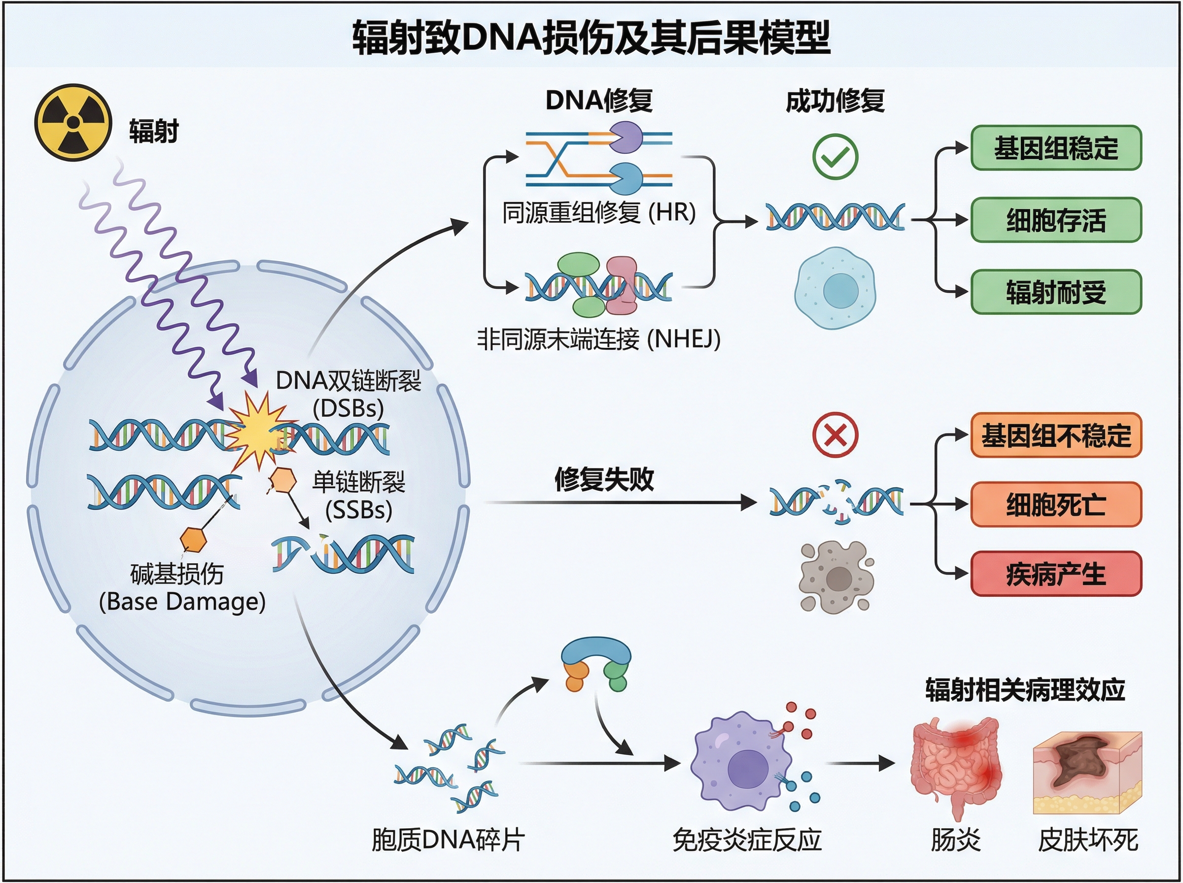 放射線誘発DNA損傷と修復経路の概要
ドラッグデリバリーと製薬イラスト
薬物の製剤化とデリバリーは主要なトピックであり、ホットメルト押出成形からナノ粒子ベースのシステムまで、あらゆるプロンプトが寄せられています。
医薬品製剤
High-resolution (300 dpi), landscape (16:9)
scientific graphical abstract for pharmaceutical research article:
"Hot Melt Extrusion-Based Solid Dispersions."
Show extrusion process, polymer-drug mixing,
amorphous solid dispersion formation,
dissolution enhancement comparison.
Clean graphical abstract style for pharmaceutical journal.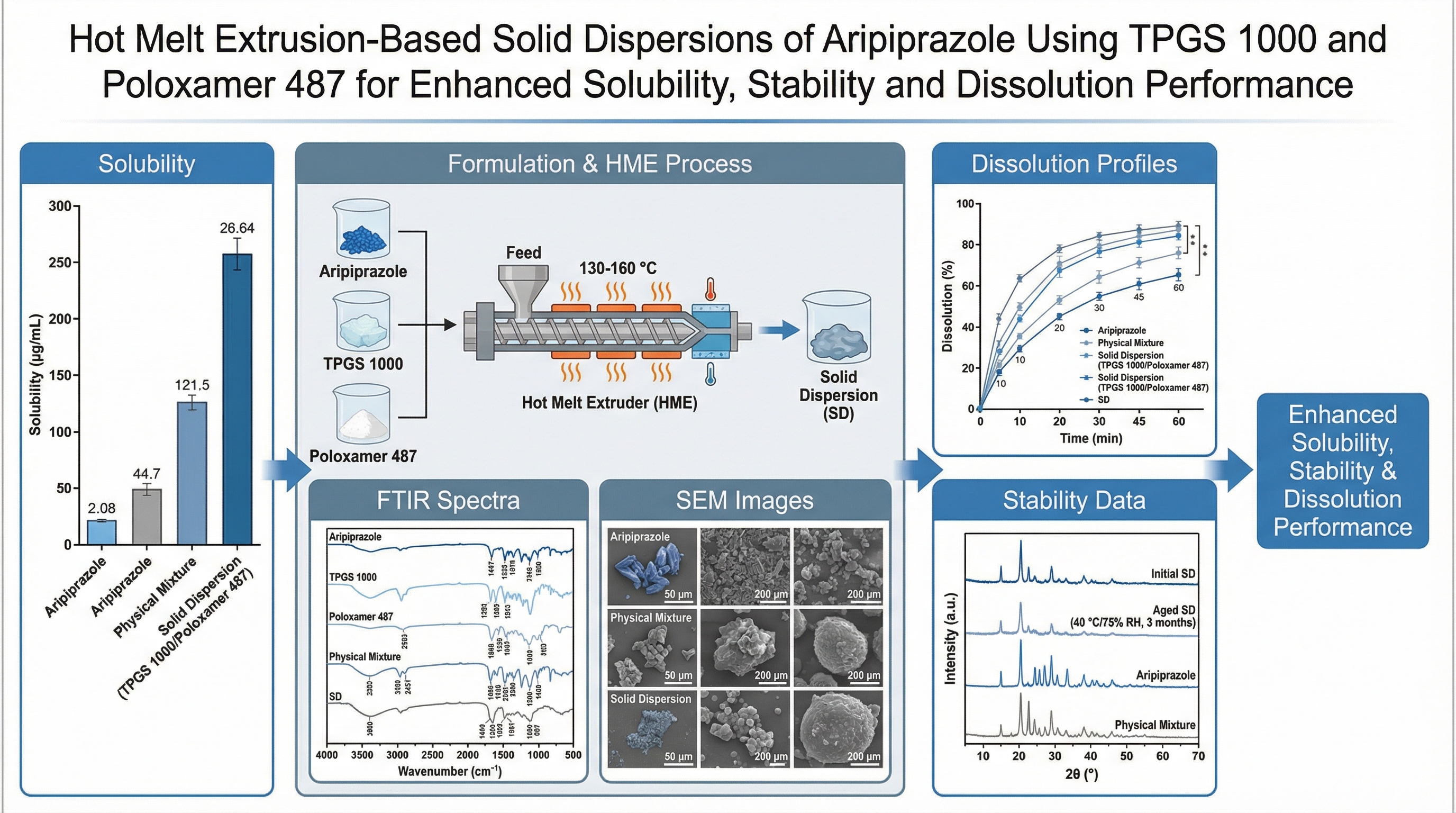 ホットメルト押出成形ベースの固体分散体のグラフィカルアブストラクト
生体医療機器のコンセプト
Academic journal-style illustration (Nature quality)
of a fully encapsulated, capsule-shaped micro-implant
with no exposed wires.
Vector-based minimalist style with detailed cross-sectional view.
Show internal electronics, biocompatible coating layers,
wireless power and data transfer components.
Suitable for biomedical engineering journal cover.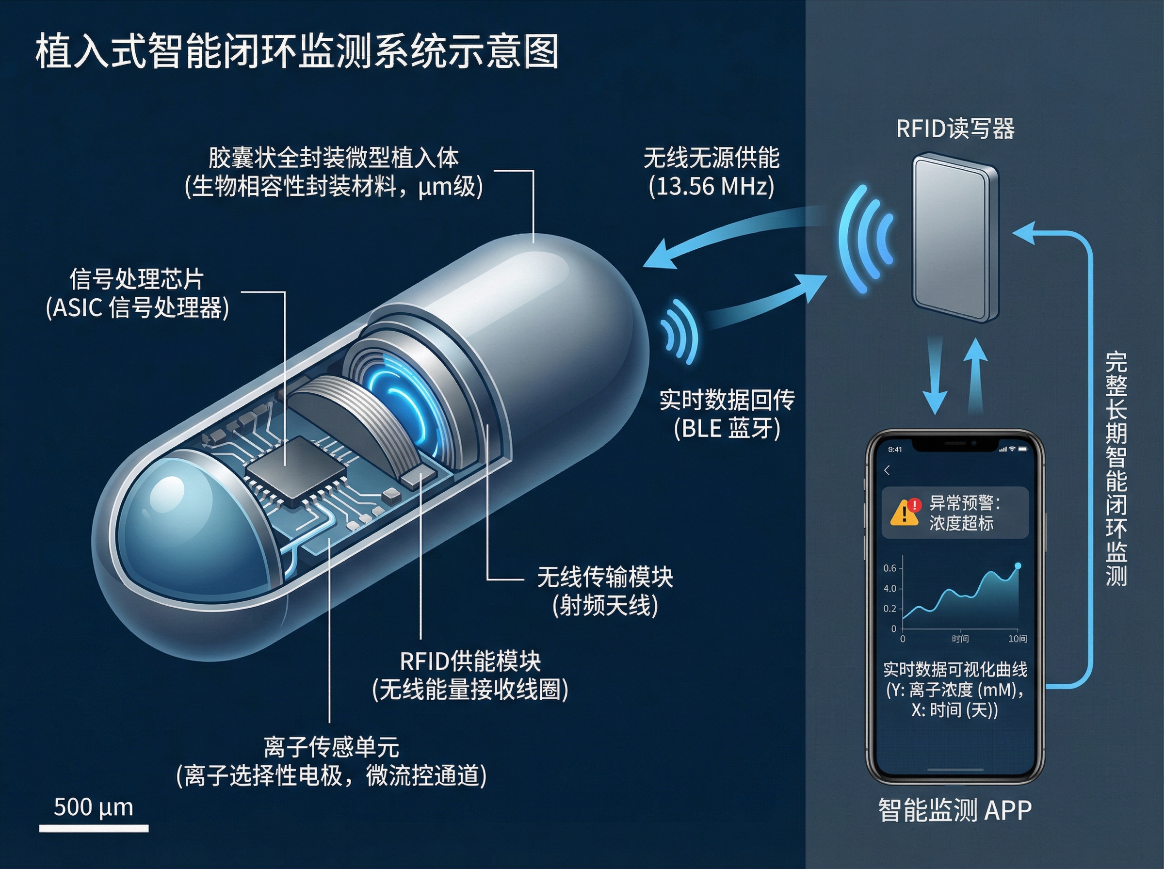 断面の詳細を示すカプセル型マイクロインプラント
動物実験の模式図
動物実験の図は、特にメソッド(手法)セクションにおいて、最も頻繁にリクエストされるイラストの一つです。
生体内(In Vivo)の手順
研究者は定期的に、標準的な実験手順のイラストを必要とします。
Generate an image of a C57 mouse undergoing gavage.
Show proper restraint technique, gavage needle insertion,
anatomical reference points.
Clean scientific illustration style,
suitable for methods section of biomedical paper.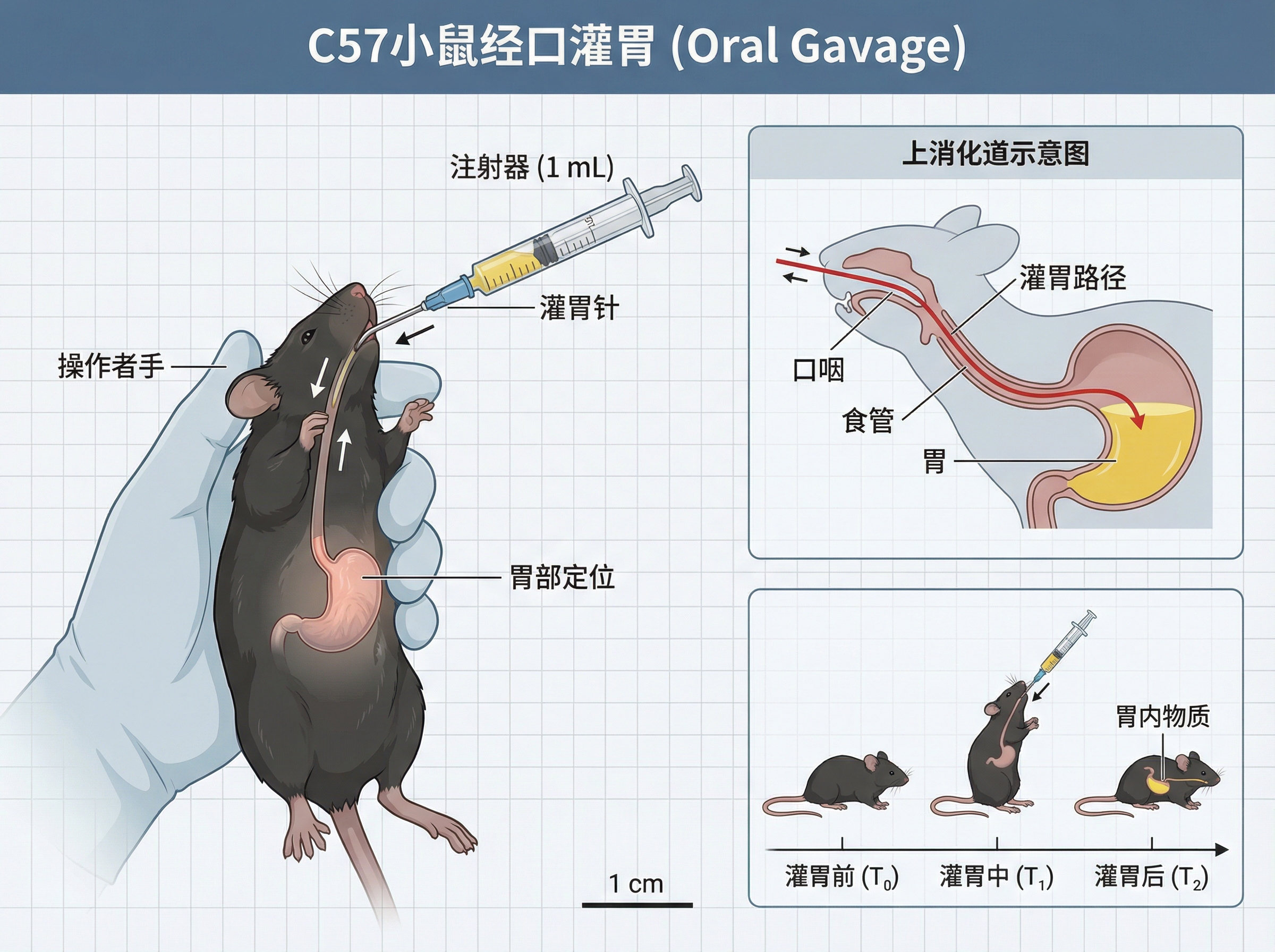 C57マウスの経口投与手順のイラスト
細胞培養プロトコル
Schematic diagram, in the style of BioRender,
illustrating the primary culture of C57 mouse astrocytes.
Show brain tissue dissection, enzymatic digestion,
cell seeding, culture medium change schedule,
passage timeline with cell morphology changes.
Step-by-step protocol diagram for neuroscience methods.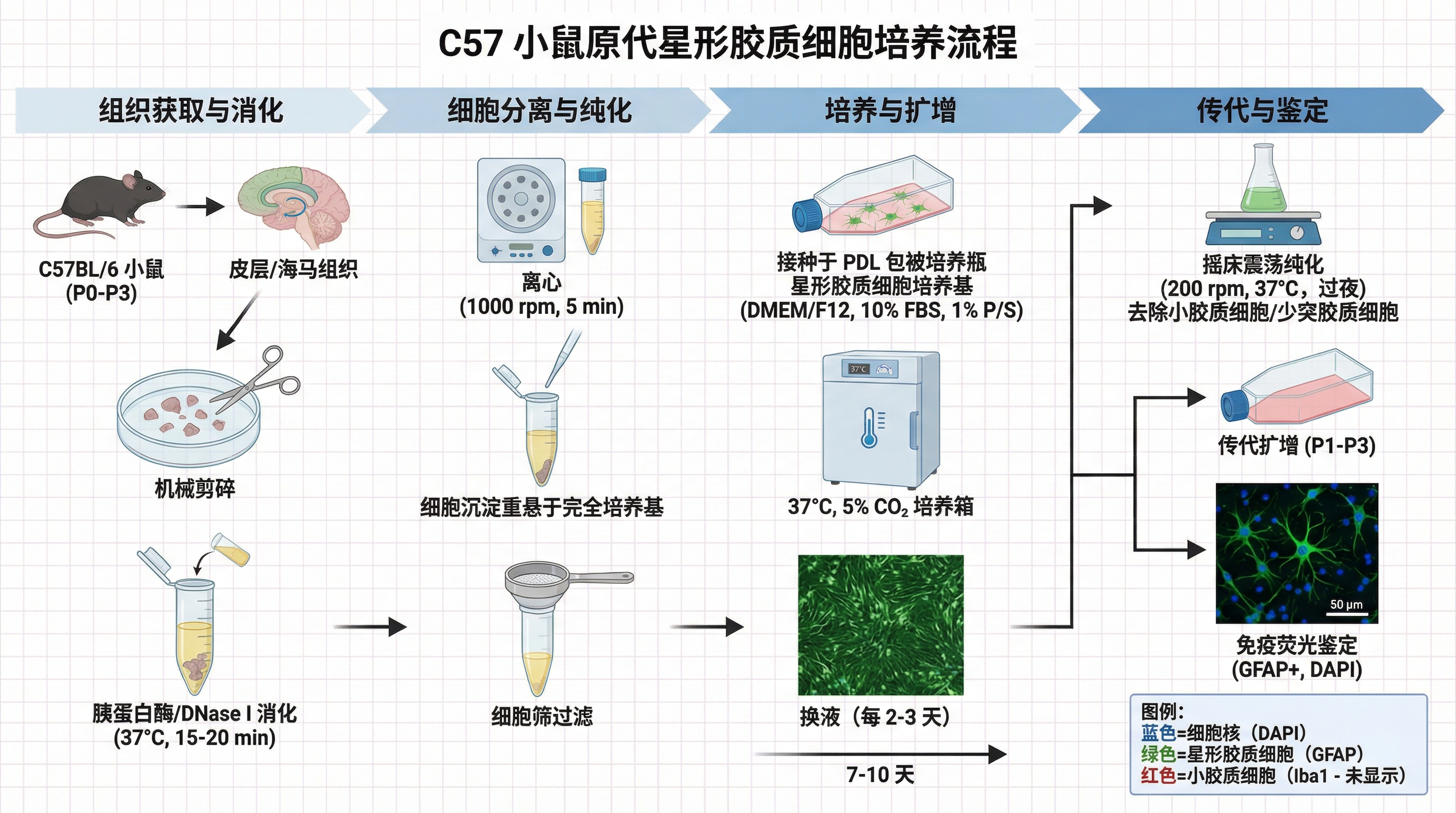 C57マウスアストロサイトの初代培養プロトコル
細胞生物学のためのプロンプト作成のヒント
成功した生物医学イラストに見られるパターンに基づき、含めるべき重要な要素を以下に示します。
必須コンポーネント
| コンポーネント | 例 | 重要な理由 |
|---|---|---|
| 細胞タイプ | "C57 mouse astrocytes" | 形態とコンテキストを決定する |
| 経路名 | "AMPK/PGC-1α cascade" | 正しい分子の登場人物を確実にする |
| コンパートメント | "cell membrane, cytoplasm, nucleus" | 空間的配置を確立する |
| 方向性 | "leading to", "resulting in" | 因果関係の流れを明確にする |
| スタイル参照 | "BioRender-style" or "Nature quality" | 視覚的な期待値を設定する |
成功するプロンプトでよく使われるキーワード
効果的な生物医学プロンプトでは、一貫して以下の用語が使用されています。
- 構造に関する言葉: "schematic diagram"(模式図), "mechanism"(メカニズム), "pathway"(経路)
- 動作に関する言葉: "illustrating"(〜を描いた), "leading to"(〜につながる), "resulting in"(〜の結果となる), "activation"(活性化)
- 具体性: 正確なタンパク質名、遺伝子名、細胞タイプ
- ターゲットとするジャーナル: "suitable for Nature/Cell/The Lancet"(Nature/Cell/The Lancetに適した)
避けるべきこと
- 曖昧な説明: 「細胞を描いて」ではなく、どの細胞タイプでどのようなプロセスなのかを指定してください。
- 方向性の欠如: 矢印や流れの方向を省略すること。
- 過密な経路: 既知のすべての相互作用ではなく、論文で扱う特定の経路に焦点を当ててください。
- 不一致な命名法: 標準的な遺伝子/タンパク質の命名規則を使用してください。
細胞生物学イラストの作成を始める
AIを使って生物医学研究の図表を一新しましょう。
- SciDraw AI Drawing にアクセス
- Mechanism Illustration(メカニズムイラスト)または Signaling Pathway(シグナル伝達経路)テンプレートを選択
- 特定の経路、細胞タイプ、分子の登場人物を記述
- 論文に使用できる出版品質の図を生成
すでにAIを活用してシグナル伝達経路図、メカニズムイラスト、実験模式図を作成している数千人の生物医学研究者の仲間に加わりましょう。
関連記事:
- メカニズム図ジェネレーター — AIでメカニズム・パスウェイ図を作成
- 細胞イラストジェネレーター — AI細胞生物学イラストツール
- Scientific Figure Generator: AI Tool for Research Papers
- TOC Graphics Generator Guide
- Thesis & Dissertation AI Diagram Guide


")
