
放射線科の学習者は、単に「きれいな画像」を求めているわけではありません。彼らが求めているのは、読影ワークフローに即した図です。つまり、所見の特定、解剖学的部位の同定、パターンの比較、そして鑑別診断のサポートができる図です。
このガイドでは、CT、MRI、胸部エックス線(CXR)の教育用ビジュアルを作成するための、再利用可能なプロンプトフレームワークを紹介します。
放射線科プロンプトの問題点
「肺結節 CT」のような短いプロンプトは、以下の要素が欠けているため、期待外れの結果に終わることがよくあります。
- 断面とビュー(軸位断/冠状断/矢状断)
- 病変の部位と境界の挙動
- 必要なラベルと矢印
- 比較対象(良性 vs 悪性、疾患A vs 疾患B)
- 教育レベル(インターン、レジデント、専門医試験対策)
画像所見のためのプロンプト構成案
Create a [CT/MRI/CXR] teaching illustration for [condition].
View: [axial/coronal/lateral/AP], with key anatomical landmarks.
Findings: [size, shape, density/signal, distribution, associated signs].
Layout: [single labeled image / side-by-side differential / progression timeline].
Labels: arrows + concise callouts + legend.
Style: clean medical textbook vector, white background, high readability.
24のハイインテント・プロンプトテンプレート
CT テンプレート
CT chest benign vs malignant lung nodule comparison, 2-panel layout.
Left: smooth margin, central calcification, stable size. Right: spiculated
margin, pleural retraction, interval growth. Add arrow callouts.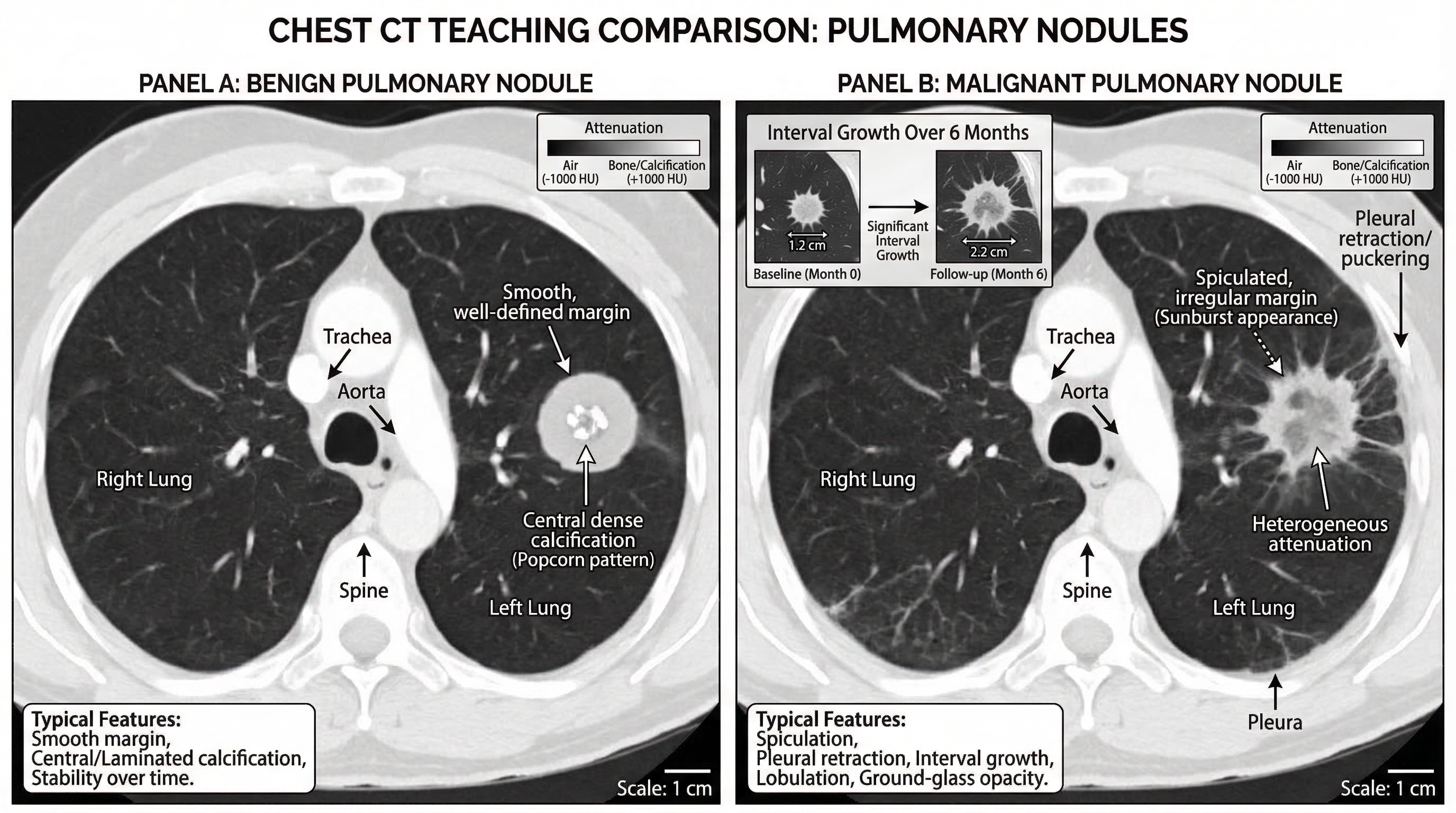
Acute appendicitis CT findings teaching figure, coronal and axial inset.
Label enlarged appendix diameter, wall thickening, periappendiceal fat stranding,
and appendicolith.
Pulmonary embolism CT angiography illustration with filling defect in segmental
artery, vessel course labeling, and right-heart strain indicators.MRI テンプレート
Acute ischemic stroke MRI sequence comparison.
DWI hyperintensity with ADC hypointensity, FLAIR mismatch concept, and lesion
territory map. Add timeline labels.
Multiple sclerosis brain MRI teaching panel with periventricular lesions,
Dawson fingers pattern, and sagittal FLAIR annotation.Pituitary adenoma MRI figure with sellar anatomy landmarks and contrast
enhancement differences vs normal pituitary tissue.CXR テンプレート
Pneumothorax CXR teaching figure with pleural line labeling,
absent peripheral lung markings, and tension signs.
Lobar pneumonia CXR map, right upper lobe consolidation with air bronchogram,
silhouette sign explanation, and differential notes.Pulmonary edema CXR pattern guide showing bat-wing opacities, Kerley B lines,
cardiomegaly, and small bilateral pleural effusion.鑑別とワークフローのテンプレート
CXR approach flowchart for dyspnea: airway, breathing, circulation,
lung fields, pleura, mediastinum, lines/tubes. Include quick red flags.
MRI ring-enhancing lesion differential chart: abscess, metastasis,
glioblastoma, toxoplasmosis. Add key discriminators in one panel.CT acute abdomen triage diagram comparing perforation, obstruction,
ischemia, and pancreatitis hallmark findings.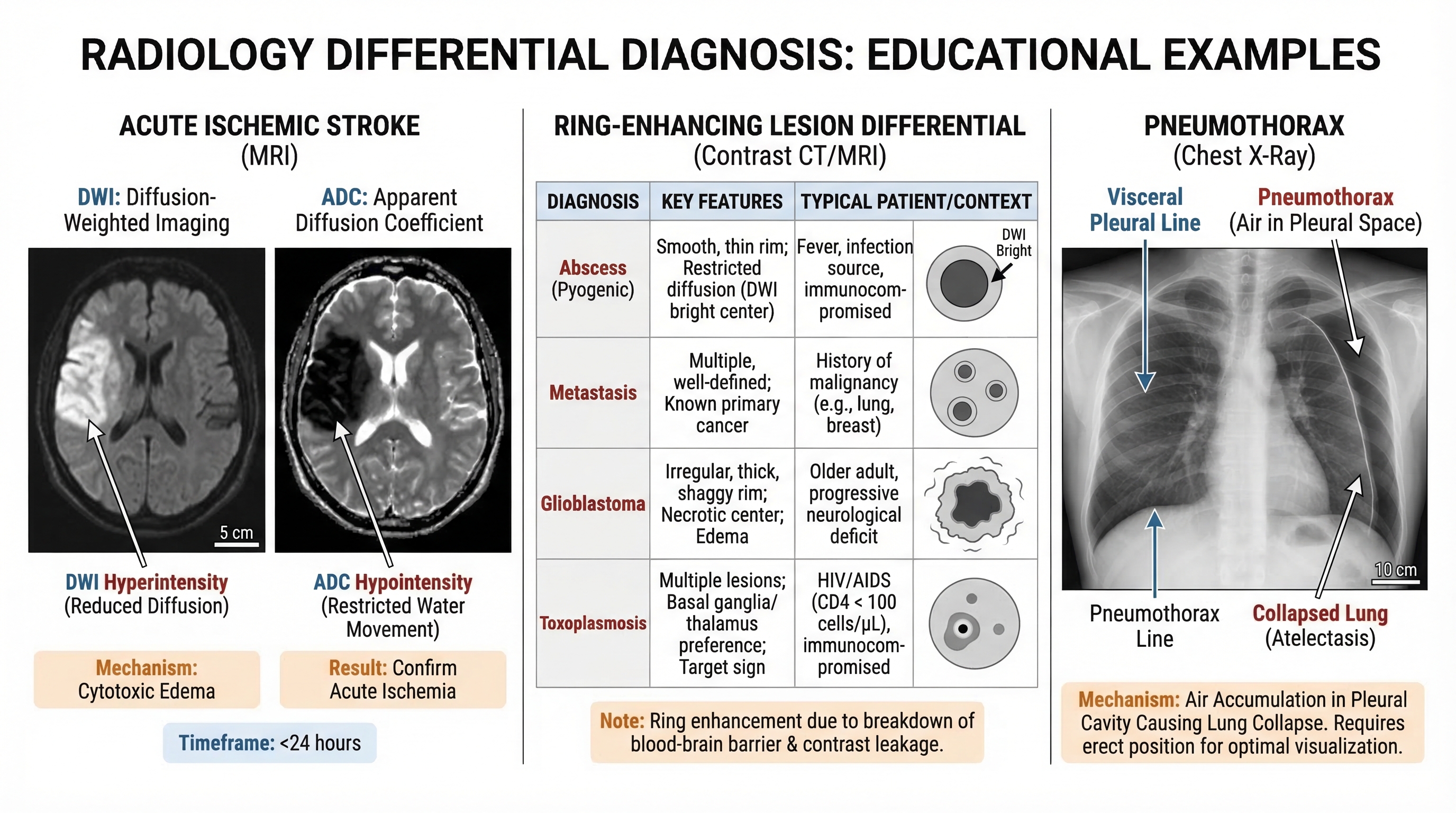
臨床的な有用性を高めるポイント
- 常に撮像面(断面)とモダリティを明示する
- 少なくとも3つの識別所見を含める
- 鑑別診断を教える際は、並列比較(side-by-side)を使用する
- ラベルは簡潔に診断的価値のあるものにし、叙述的な説明は避ける
- 論文としての洗練さよりも、まずはスライドでの読みやすさを重視して設計する
書き出し前のチェックリスト
- モダリティとシーケンスが明示されているか
- 解剖学的指標(ランドマーク)が存在するか
- 病変のコールアウト(引き出し線)が明確か
- 鑑別のヒントが一目でわかるか
- プレゼンテーションモードでも図が読みやすいか
より速く作成し、より良く教え、画像教育資産を標準化しましょう。


