Eine biomedizinische Abbildung für ein Paper, einen Förderantrag oder einen Vortrag zu erstellen, war bis vor Kurzem eine Entweder-oder-Sache: Entweder Sie beherrschten Illustrator, oder Sie bezahlten jemanden fürs Zeichnen. Heute beschreiben Sie in einem Absatz, was Sie brauchen, und die KI liefert eine Abbildung fast auf Publikationsniveau. Die Schwierigkeit hat sich von „die Software beherrschen" zu „den Prompt schreiben" verschoben.
Dieser Leitfaden bündelt 50 Prompts aus echten Forschungsprojekten, geordnet nach Einsatz (Zellbiologie, Molekularbiologie, Immunologie, medizinische Abbildungen). Kopieren, etwas anpassen — fertig.
Was Sie in diesem Leitfaden finden:
- Gebrauchsfertige Prompt-Vorlagen für gängige biomedizinische Diagramme
- Reale Beispiele aus veröffentlichten Forschungsprojekten
- Tipps zur Anpassung von Prompts an Ihre spezifischen Bedürfnisse
- Best Practices für publikationsreife Ergebnisse
1. Zellbiologie & Molekulare Mechanismen
1.1 Proteinstruktur und -funktion
Prompt: p53 DNA Binding Mechanism
The p53 protein binds to the DNA double helix via its central DNA binding domain.
Create a molecular illustration showing the tetrameric p53 structure engaging with
the major groove of DNA, highlighting the zinc coordination sites and key amino
acid residues involved in sequence-specific recognition.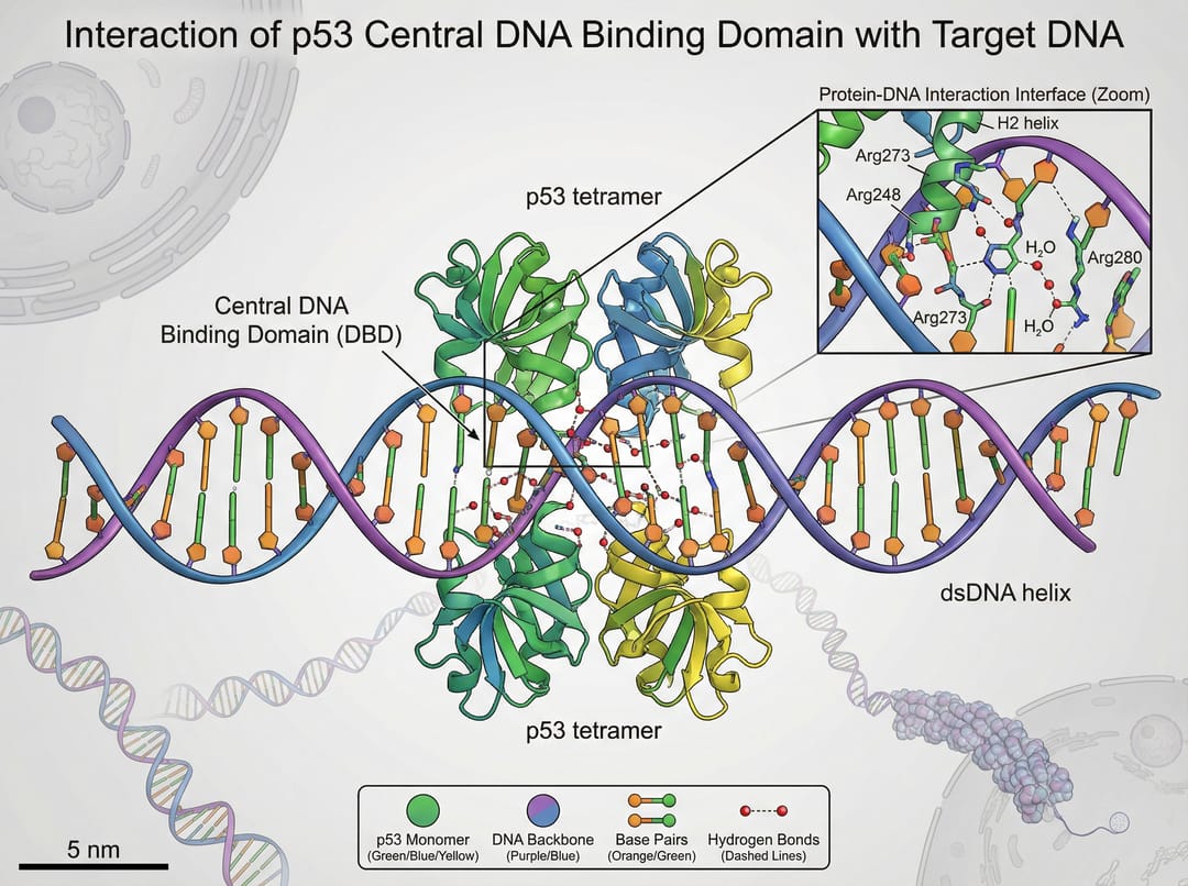
Prompt: ECM Proteomics in Cardiovascular Disease
"Applications of ECM-specific proteomics in cardiovascular disease".
The figure should be vertically structured, showing the workflow from tissue
sample collection through mass spectrometry analysis to biomarker discovery,
with emphasis on extracellular matrix protein identification.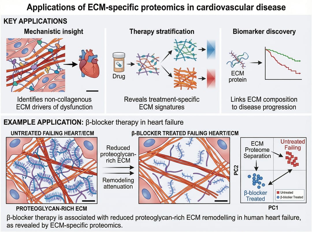
1.2 Zelluläre Prozesse
Prompt: Drug Delivery Mechanism - FlaTox System
Create a simplified schematic illustrating the FlaTox-mediated delivery of
flagellin into intestinal epithelial cells. Show the protective antigen (PA)
binding to cell surface receptors, pore formation, and subsequent translocation
of the flagellin fusion protein into the cytoplasm where it activates NLRC4
inflammasome signaling.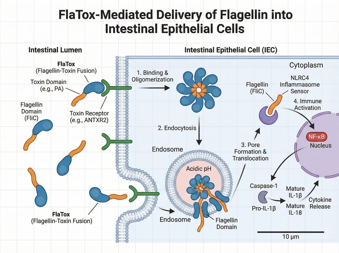
Prompt: Postprandial Glucose Effects
Illustration depicting the postprandial glucose elevation following a high-sugar
meal and its subsequent detrimental effects on vascular endothelium. Show the
timeline from meal ingestion through glucose spike to endothelial dysfunction,
including oxidative stress markers and inflammatory pathway activation.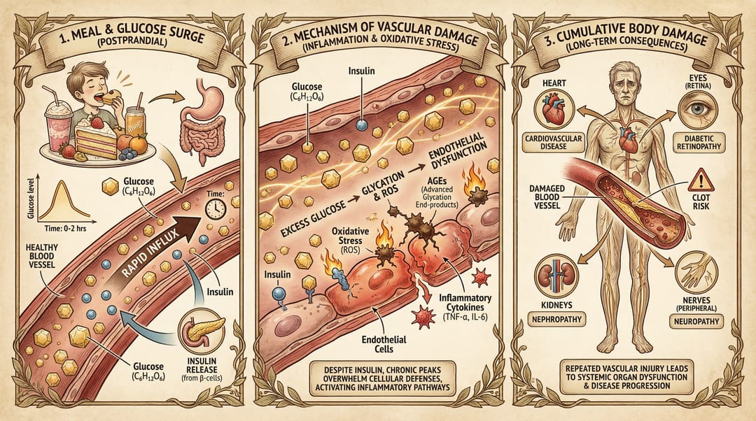
2. Immunologie & Krankheitsforschung
2.1 Immunzellstudien
Prompt: NK Cell Homeostasis in Autism Research
This study draws the following key conclusions:
Mice with 15q duplication autism exhibit significant NK cell homeostasis
imbalance. Create a figure showing terminally mature NK cells, immature NK cell
populations, and the disrupted balance between them, with quantitative markers
and statistical significance indicators.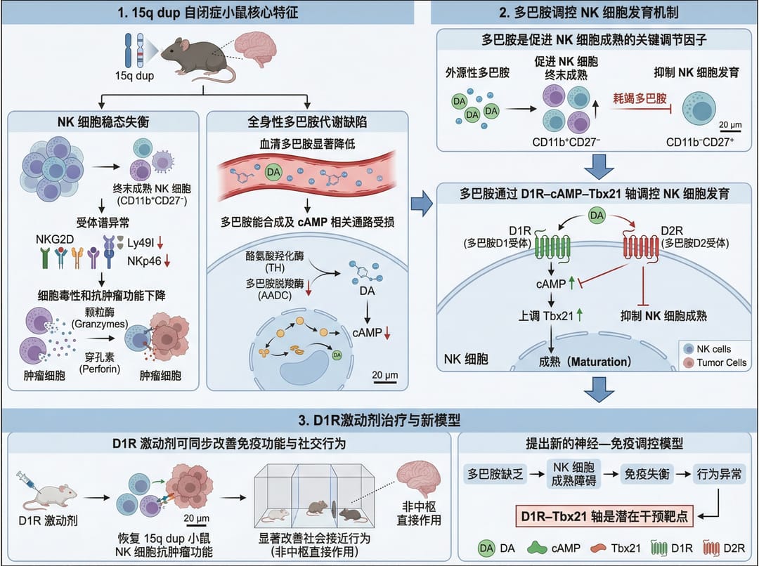
2.2 Epidemiologie & Globale Gesundheit
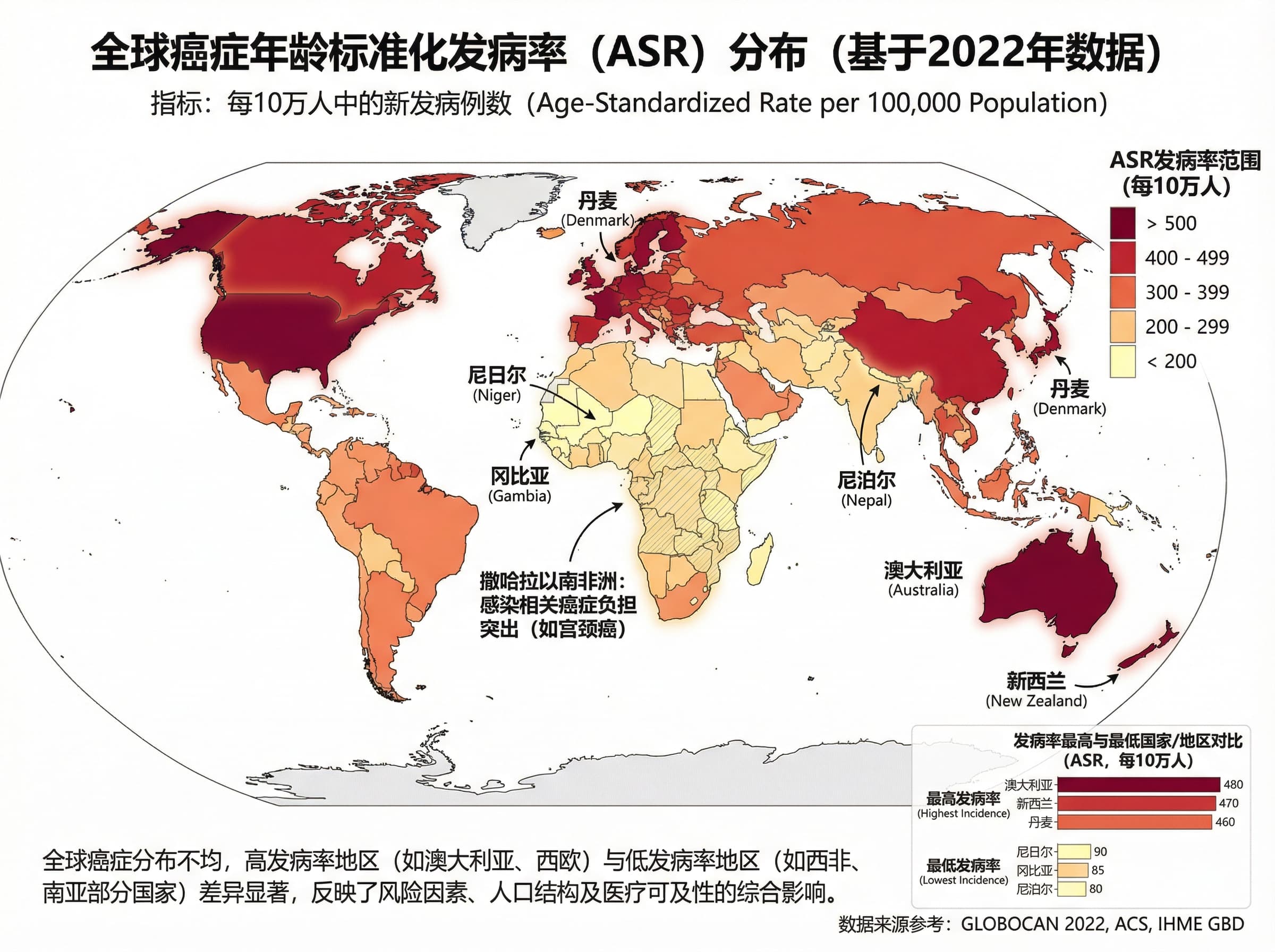
Prompt: Global Cancer Incidence Map
Core Theme: To create a world map illustrating the global distribution of
age-standardized cancer incidence rates (ASR). Use a choropleth map with
gradient colors from light (low incidence) to dark (high incidence), include
regional labels, and add a color legend with ASR ranges per 100,000 population.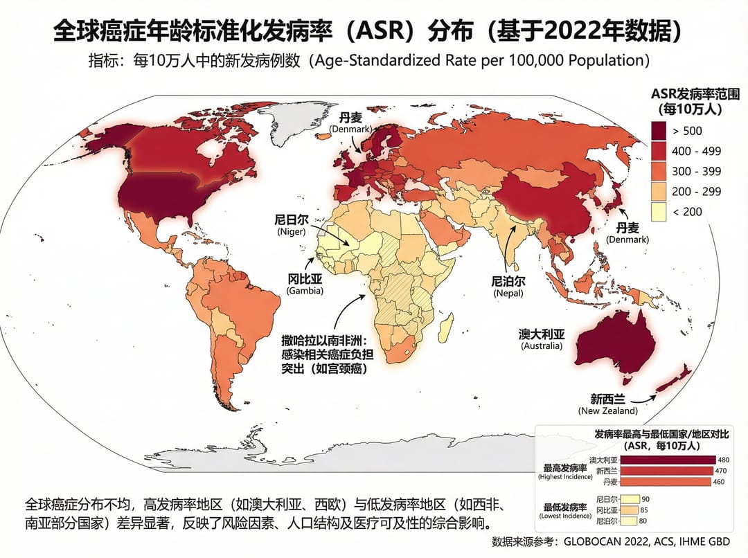
3. Laborprotokolle & Methoden
3.1 Workflows der Proteinanalyse
Prompt: SDS-PAGE Protocol
Here is a protocol for protein extraction, denaturation, gel preparation, and
sample loading for SDS-PAGE. Create a step-by-step workflow diagram showing:
(1) Total protein extraction from tissue samples
(2) BCA protein quantification
(3) Sample preparation with loading buffer
(4) Gel electrophoresis setup
(5) Coomassie staining and imaging
Include timing annotations and key reagent concentrations.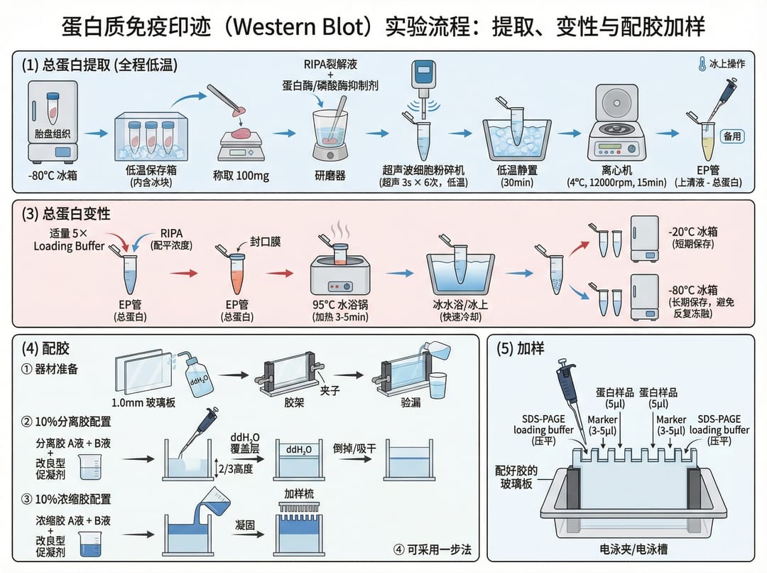
Prompt: Immunohistochemistry Flowchart
An immunohistochemistry (IHC) flowchart:
(1) Preparation of paraffin sections:
① Tissue fixation
② Dehydration and embedding
③ Sectioning at 4μm thickness
(2) Antigen retrieval (heat-induced or enzymatic)
(3) Blocking and primary antibody incubation
(4) Secondary antibody and DAB development
(5) Counterstaining and mounting
Use a vertical flow with icons for each step.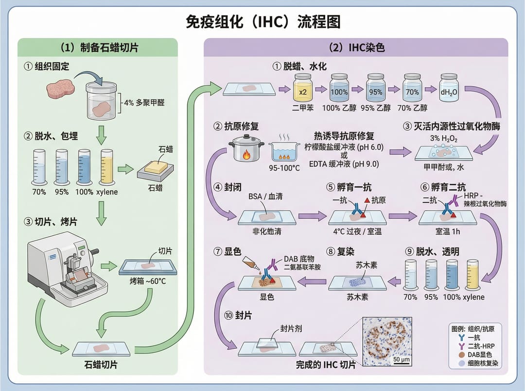
3.2 Methoden für systematische Übersichtsarbeiten
Prompt: PRISMA 2020 Flow Diagram
Create a PRISMA 2020 flow diagram as a vertical process chart with a 4:3 aspect
ratio. Employ a clean, professional style with rectangular boxes connected by
arrows. Include four main sections: Identification (database searches, n=XXX),
Screening (title/abstract review, full-text assessment), Eligibility (inclusion/
exclusion criteria applied), and Included (final studies for synthesis). Add
boxes for duplicates removed and reasons for exclusion at each stage.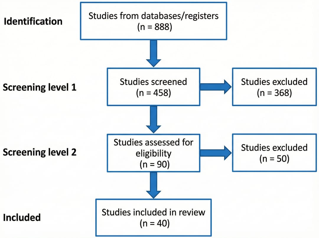
4. Klinische & Medizinische Illustrationen
4.1 Geburtshilfe & Gynäkologie
Prompt: Amniotic Membrane Rupture Types
Medical illustration depicting two distinct types of amniotic membrane rupture:
(1) A high leak, characterized by a small perforation in the upper portion of
the amniotic sac with gradual fluid loss
(2) A complete rupture at the cervical os with sudden, large-volume fluid
discharge
Show anatomical cross-sections with labeled structures including uterus,
placenta, amniotic fluid, and fetal position.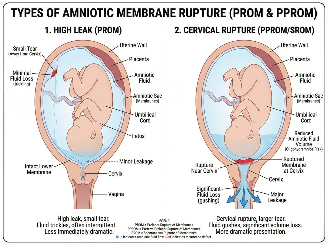
5. Best Practices für das Schreiben von Prompts
Strukturieren Sie Ihre Prompts effektiv
Basierend auf der Analyse von 645 erfolgreichen biomedizinischen Prompts sind dies die Schlüsselelemente:
1. Beginnen Sie mit dem Kontext
- Geben Sie die Art der Illustration an (Schema, Flussdiagramm, Mechanismus-Diagramm)
- Geben Sie die Zielgruppe an (Journaleinreichung, Präsentation, Förderantrag)
2. Integrieren Sie technische Details
- Benennen Sie spezifische Proteine, Signalwege oder biologische Strukturen
- Verwenden Sie die korrekte wissenschaftliche Nomenklatur
- Geben Sie Maßstab oder Vergrößerung an, falls relevant
3. Beschreiben Sie visuelle Anforderungen
- Seitenverhältnis (4:3 Querformat, 3:4 Hochformat, 16:9 Breitbild)
- Bevorzugte Farbschemata
- Platzierung von Beschriftungen und Schriftstil
- Verbindungspfeile und Flussrichtung
4. Beziehen Sie sich auf Publikationsstandards
- Erwähnen Sie den Stil des Zieljournals, falls zutreffend
- Geben Sie Anforderungen an die Auflösung an
- Beachten Sie Barrierefreiheit (farbenblindenfreundliche Paletten)
Gängige Prompt-Muster
| Anwendung | Wichtige zu enthaltene Elemente |
|---|---|
| Mechanismus-Diagramme | Molekulare Komponenten, Interaktionspfeile, zeitliche Abfolge |
| Protokoll-Workflows | Schrittnummern, Zeitangaben, Reagenzienkonzentrationen, Gerätesymbole |
| Anatomische Illustrationen | Gewebeschichten, beschriftete Strukturen, Querschnitt vs. Oberflächenansicht |
| Datenvisualisierung | Achsenbeschriftungen, Legende, statistische Annotationen, Stichprobengrößen |
| Vergleichende Abbildungen | Side-by-Side-Layout, konsistentes Styling, Hervorhebung von Unterschieden |
Beginnen Sie mit der Erstellung Ihrer biomedizinischen Illustrationen
Bereit, professionelle wissenschaftliche Abbildungen zu erstellen? Probieren Sie diese Prompts mit den KI-gestützten Illustrationswerkzeugen von SciDraw AI aus:
- Klinische-Illustrations-Generator — Medizinische und anatomische Illustrationen mit KI erstellen
- Zellillustrations-Generator — KI-gestützte Zellbiologie-Diagramme
- Erstellen Sie Ihre erste Illustration →
- Weitere Beispiele in der Galerie durchsuchen →
Alle Prompts in diesem Leitfaden stammen aus realen Forschungsprojekten und wurden auf publikationswürdige Ergebnisse getestet. Passen Sie diese an Ihre spezifischen Forschungsbedürfnisse an und beginnen Sie noch heute mit der Erstellung professioneller biomedizinischer Illustrationen.