
放射科学习者不需要随机的“漂亮图片”。他们需要符合临床阅片工作流的插图:识别征象、解剖定位、模式对比以及支持鉴别诊断。
本指南为你提供了一个可复用的提示词(Prompt)框架,用于生成 CT、MRI 和胸部 X 光(CXR)的教学视觉资料。
放射科提示词的常见问题
像“肺结节 CT”这样简短的提示词往往会失败,因为它们忽略了:
- 切面和视图(轴状位/冠状位/矢状位)
- 病变位置和边界特征
- 必要的标签和箭头
- 对比目标(良性 vs 恶性,疾病 A vs 疾病 B)
- 教育水平(实习生、住院医、专科考试准备)
影像征象提示词公式
Create a [CT/MRI/CXR] teaching illustration for [condition].
View: [axial/coronal/lateral/AP], with key anatomical landmarks.
Findings: [size, shape, density/signal, distribution, associated signs].
Layout: [single labeled image / side-by-side differential / progression timeline].
Labels: arrows + concise callouts + legend.
Style: clean medical textbook vector, white background, high readability.
24 个高针对性提示词模板
CT 模板
CT chest benign vs malignant lung nodule comparison, 2-panel layout.
Left: smooth margin, central calcification, stable size. Right: spiculated
margin, pleural retraction, interval growth. Add arrow callouts.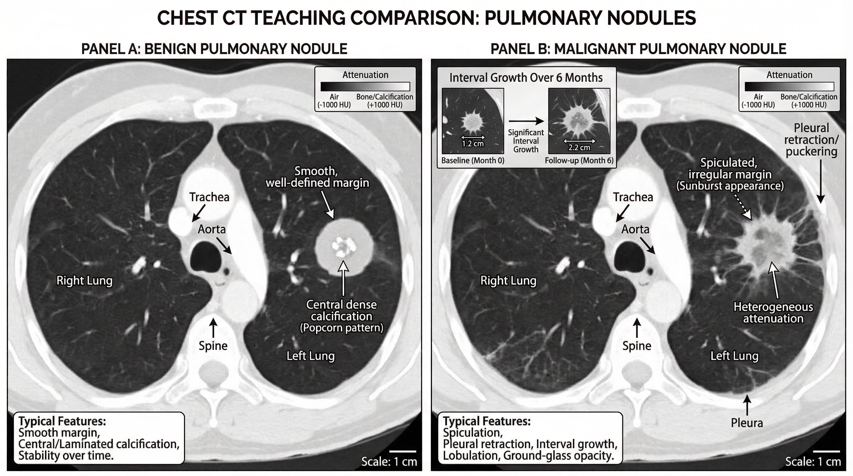
Acute appendicitis CT findings teaching figure, coronal and axial inset.
Label enlarged appendix diameter, wall thickening, periappendiceal fat stranding,
and appendicolith.
Pulmonary embolism CT angiography illustration with filling defect in segmental
artery, vessel course labeling, and right-heart strain indicators.MRI 模板
Acute ischemic stroke MRI sequence comparison.
DWI hyperintensity with ADC hypointensity, FLAIR mismatch concept, and lesion
territory map. Add timeline labels.
Multiple sclerosis brain MRI teaching panel with periventricular lesions,
Dawson fingers pattern, and sagittal FLAIR annotation.Pituitary adenoma MRI figure with sellar anatomy landmarks and contrast
enhancement differences vs normal pituitary tissue.CXR 模板
Pneumothorax CXR teaching figure with pleural line labeling,
absent peripheral lung markings, and tension signs.
Lobar pneumonia CXR map, right upper lobe consolidation with air bronchogram,
silhouette sign explanation, and differential notes.Pulmonary edema CXR pattern guide showing bat-wing opacities, Kerley B lines,
cardiomegaly, and small bilateral pleural effusion.鉴别诊断与工作流模板
CXR approach flowchart for dyspnea: airway, breathing, circulation,
lung fields, pleura, mediastinum, lines/tubes. Include quick red flags.
MRI ring-enhancing lesion differential chart: abscess, metastasis,
glioblastoma, toxoplasmosis. Add key discriminators in one panel.CT acute abdomen triage diagram comparing perforation, obstruction,
ischemia, and pancreatitis hallmark findings.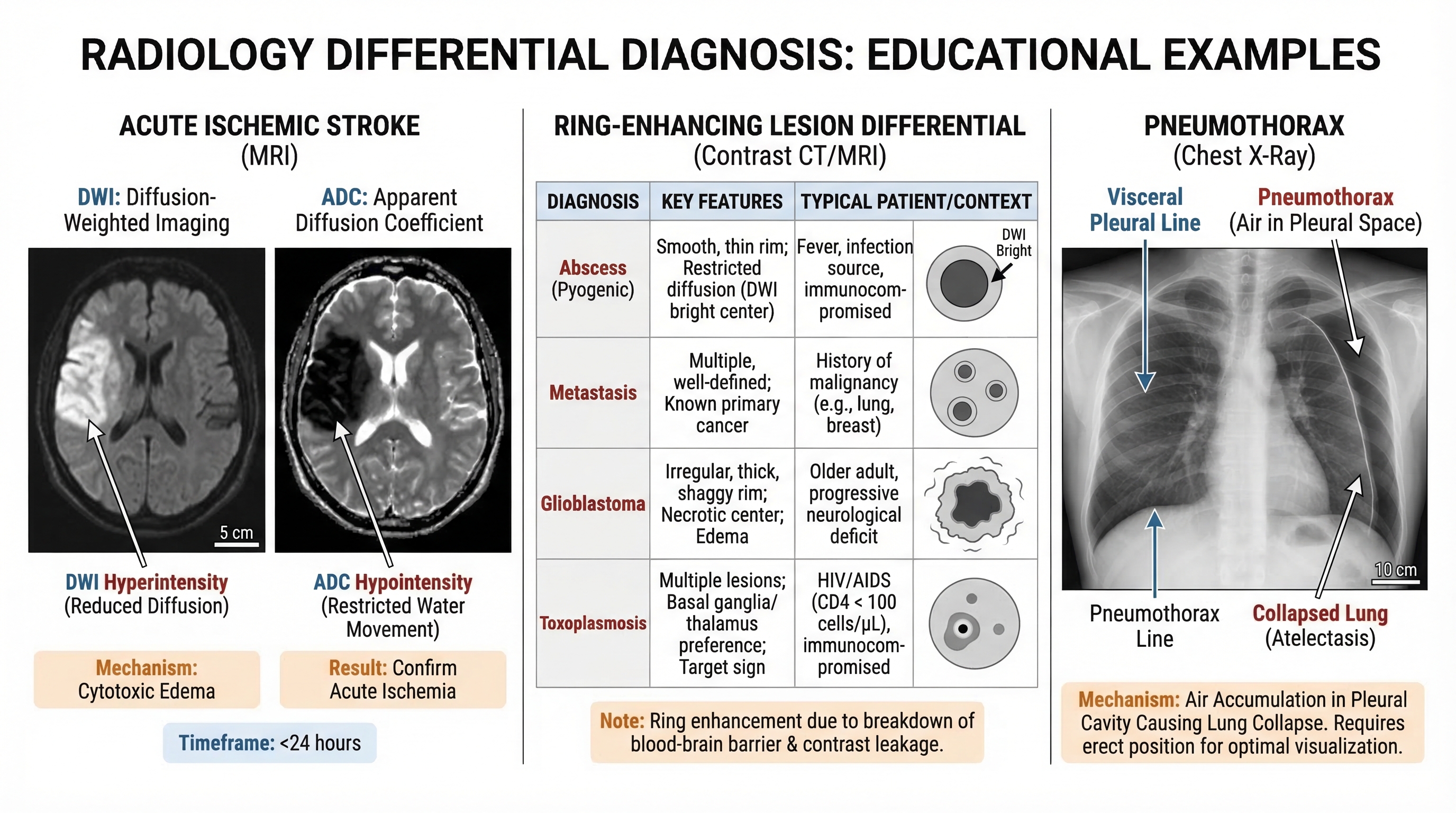
如何提升临床实用性
- 始终明确影像切面和检查技术(Modality)
- 至少包含 3 个具有鉴别意义的征象
- 在教学鉴别诊断时,使用并排对比布局
- 标签应保持简短且具有诊断性,而非叙述性
- 设计时优先考虑幻灯片的可读性,其次才是期刊级的精细度
导出前检查清单
- 检查技术和序列已明确命名
- 包含解剖学标志
- 病变标注清晰无歧义
- 鉴别诊断线索一目了然
- 插图在演示模式下依然清晰可读
更快地构建、更好地教学,并标准化你的影像教育资产。


