
영상의학 학습자들에게는 단순히 무작위로 생성된 "예쁜 이미지"가 필요한 것이 아닙니다. 그들에게는 판독 워크플로우에 부합하는 그림이 필요합니다: 소견 식별, 해부학적 위치 파악, 패턴 비교, 그리고 감별 진단 지원.
이 가이드는 CT, MRI, 흉부 엑스레이(CXR) 교육용 시각 자료를 위한 재사용 가능한 프롬프트 프레임워크를 제공합니다.
영상의학 프롬프트의 문제점
"lung nodule CT(폐 결절 CT)"와 같은 짧은 프롬프트는 다음과 같은 요소들이 누락되어 있어 실패하는 경우가 많습니다:
- 단면 및 뷰 (축상면/관상면/시상면)
- 병변 위치 및 경계 특성
- 필수 레이블 및 화살표
- 비교 대상 (양성 vs 악성, 질환 A vs B)
- 교육 수준 (인턴, 레지던트, 전문의 시험 준비)
영상 소견을 위한 프롬프트 공식
Create a [CT/MRI/CXR] teaching illustration for [condition].
View: [axial/coronal/lateral/AP], with key anatomical landmarks.
Findings: [size, shape, density/signal, distribution, associated signs].
Layout: [single labeled image / side-by-side differential / progression timeline].
Labels: arrows + concise callouts + legend.
Style: clean medical textbook vector, white background, high readability.
24가지 고의도(High-Intent) 프롬프트 템플릿
CT 템플릿
CT chest benign vs malignant lung nodule comparison, 2-panel layout.
Left: smooth margin, central calcification, stable size. Right: spiculated
margin, pleural retraction, interval growth. Add arrow callouts.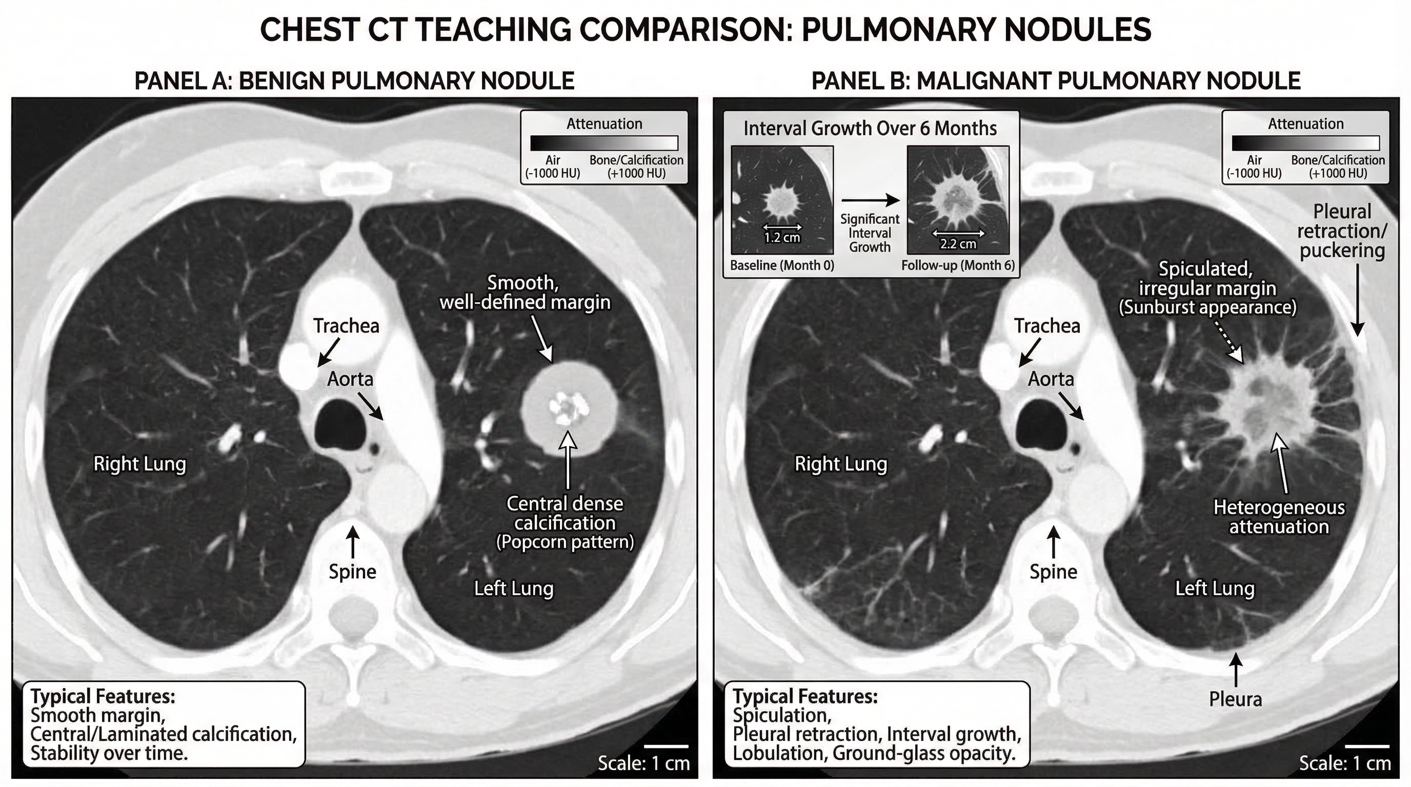
Acute appendicitis CT findings teaching figure, coronal and axial inset.
Label enlarged appendix diameter, wall thickening, periappendiceal fat stranding,
and appendicolith.
Pulmonary embolism CT angiography illustration with filling defect in segmental
artery, vessel course labeling, and right-heart strain indicators.MRI 템플릿
Acute ischemic stroke MRI sequence comparison.
DWI hyperintensity with ADC hypointensity, FLAIR mismatch concept, and lesion
territory map. Add timeline labels.
Multiple sclerosis brain MRI teaching panel with periventricular lesions,
Dawson fingers pattern, and sagittal FLAIR annotation.Pituitary adenoma MRI figure with sellar anatomy landmarks and contrast
enhancement differences vs normal pituitary tissue.CXR 템플릿
Pneumothorax CXR teaching figure with pleural line labeling,
absent peripheral lung markings, and tension signs.
Lobar pneumonia CXR map, right upper lobe consolidation with air bronchogram,
silhouette sign explanation, and differential notes.Pulmonary edema CXR pattern guide showing bat-wing opacities, Kerley B lines,
cardiomegaly, and small bilateral pleural effusion.감별 진단 및 워크플로우 템플릿
CXR approach flowchart for dyspnea: airway, breathing, circulation,
lung fields, pleura, mediastinum, lines/tubes. Include quick red flags.
MRI ring-enhancing lesion differential chart: abscess, metastasis,
glioblastoma, toxoplasmosis. Add key discriminators in one panel.CT acute abdomen triage diagram comparing perforation, obstruction,
ischemia, and pancreatitis hallmark findings.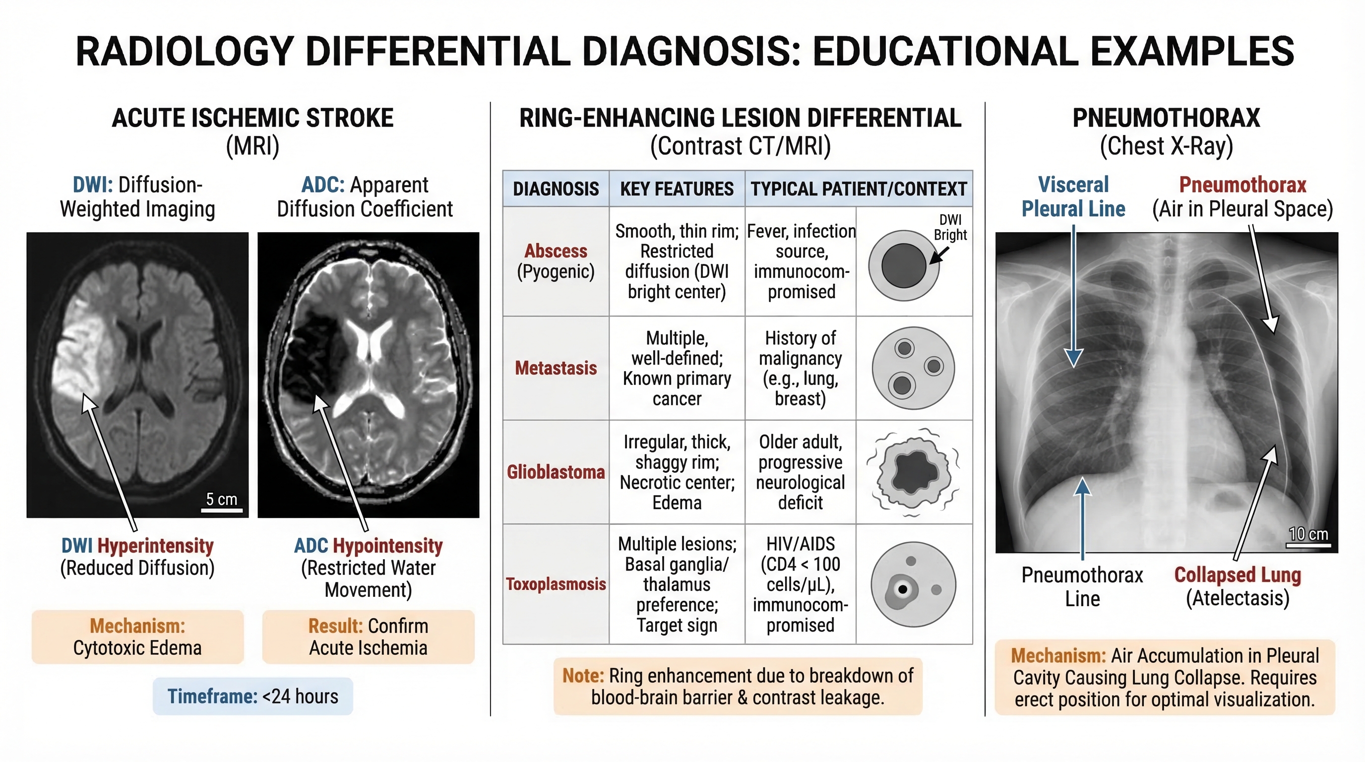
임상적 유용성을 높이는 방법
- 항상 영상 단면(Plane)과 모달리티(Modality)를 정의하십시오.
- 최소 3가지 이상의 변별력 있는 소견을 포함하십시오.
- 감별 진단을 가르칠 때는 병렬 비교(Side-by-side comparison)를 활용하십시오.
- 레이블은 서술형이 아닌, 짧고 진단적인 용어로 유지하십시오.
- 논문용 정교함보다는 슬라이드 가독성을 우선하여 디자인하십시오.
최종 점검 리스트
- 모달리티와 시퀀스가 명확하게 명시되었는가
- 해부학적 지표가 포함되었는가
- 병변 콜아웃이 모호하지 않은가
- 감별 포인트가 한눈에 들어오는가
- 발표 모드에서도 그림의 가독성이 유지되는가
더 빠르게 제작하고, 더 효과적으로 가르치며, 영상 교육 자산을 표준화하십시오.


